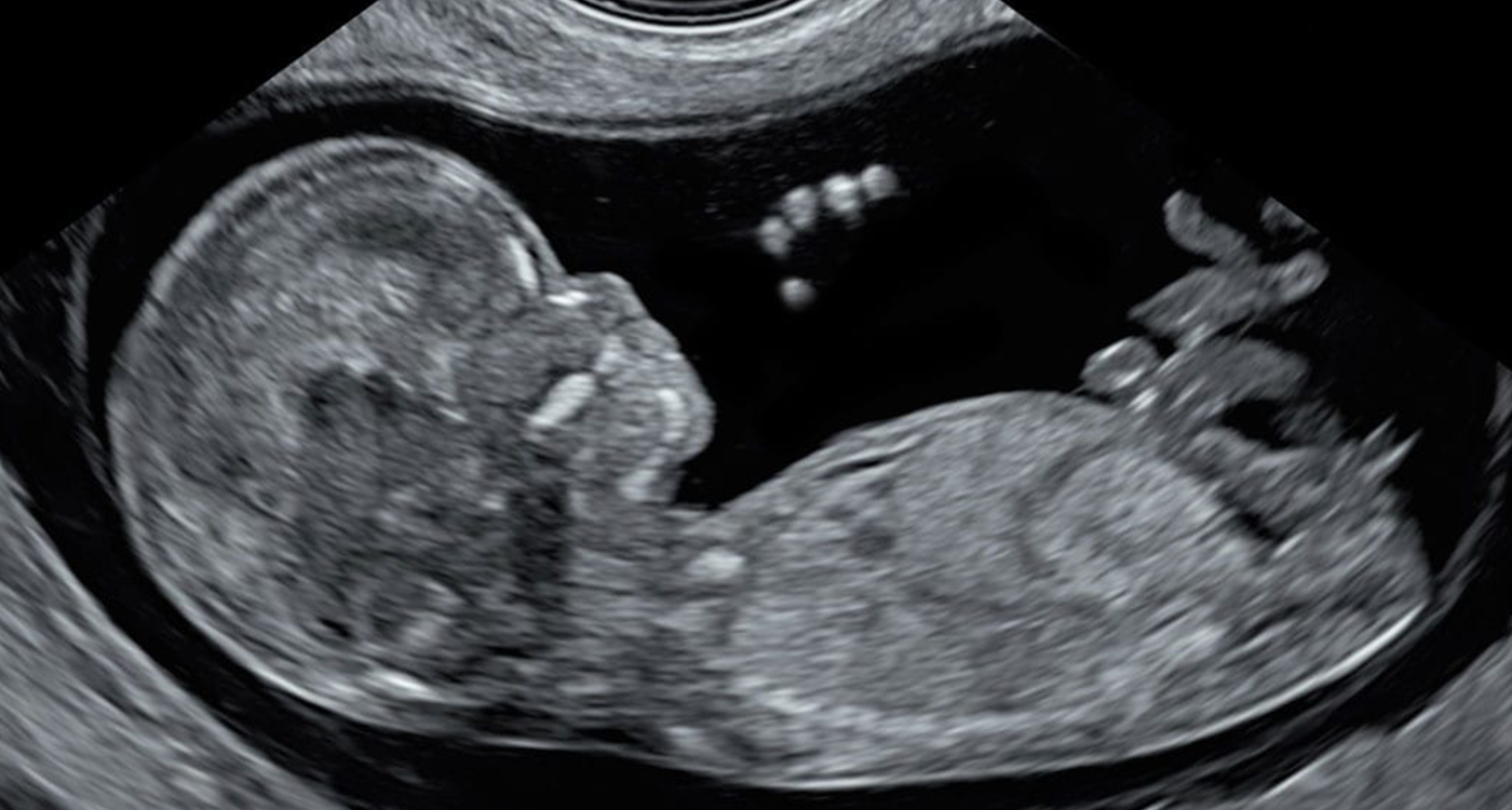
Cardiac Scan
We believe that the heart of every baby must be carefully examined and we perform Fetal Echocardiography for free for all babies we scan

Why is it important to examine the baby’s heart during every scan?
Heart defects (cardiac anomalies or congenital heart disease) are the most common human anomalies. In fact, cardiac anomalies are much more common than trisomy 21 (Down’s Syndrome) and can sometimes be life-threatening to the baby.
The vast majority of heart defects affect babies that have healthy parents with no family history of cardiac problems; however, in some cases, maternal diabetes or increased nuchal translucency (NT) can suggest and increased risk of cardiac anomaly.
Key facts about the baby’s heart:
- Composed of four chambers, four valves, a foramen ovale, two arteries, and six veins,
- Relatively small, occupying about a third of the baby’s chest,
- The heart rate is about 2.5 contractions per second
The complexity of the baby’s heart in combination with the above-mentioned descriptions, makes imaging very challenging. Furthermore, heart defects are highly variable, with over 100 heart anomalies identified to date. This highlights why many heart defects (even life-threatening ones) often remain undiagnosed until birth. Currently, the national target (NHS) for detection of severe heart defects by antenatal screening is 50%. This means that every second baby with a severe heart disease that has needed cardiac surgery within the first year of life, has been delivered without a prenatal diagnosis.
My baby has a normal heartbeat. Does this mean all serious heart anomalies can be excluded?
It is often misunderstood that a normal fetal heartbeat excludes all structural anomalies. While it may sound surprising, the vast majority of babies born with severe and even lethal heart defects have completely normal heart beats. An examination of the heart during pregnancy screening is mostly focused on examining its structure.
In some rare cases, the baby may have an abnormal heart rhythm (irregular, very slow, very high). These cases would be monitored and treated by specialist fetal/paediatric cardiologists.
We do a thorough scan of the heart during any of our scans performed from 12-36 weeks.
Not only are we experts in performing fetal echocardiography or fetal echo (examination of your baby’s heart), we also have a long-standing record of training other professionals in the technique.
We have developed a three-stage protocol for congenital heart disease screening:
- Early Baby Scan at 12-13 weeks: By this stage of pregnancy we are able to exclude some severe heart defects, such as transposition of the great arteries and hypoplastic left heart syndrome, as well as provide reassurance for the parents.
- Anomaly Scan at 21-23 weeks: By this stage we are able to exclude some major anomalies which would have been undetectable at 12-13 weeks. We will also try to exclude some moderate and minor cardiac defects such as ventricular septal defects (“hole in the heart”).
- Baby Development Scan at 28-34 weeks: During this scan we aim to exclude heart anomalies, which manifest later in the pregnancy. These conditions are rare; however, some of them can be very serious or even life-threatening. These include heart rhythm abnormalities, heart tumours, heart valve abnormalities, and some ventricular septal defects may only be detectable at this stage.
Unfortunately, it is impossible to exclude all heart defects in 100% of pregnancies because some anomalies may manifest only after birth. These include coarctation of the aorta, patent ductus arteriosus, among others. In other rare circumstances, detectable cardiac anomalies go undiagnosed due to technical reasons during imaging.
Our checklist for baby’s heart examination (Fetal Echo) includes:
At 12-13 weeks the fetal heart is about the size of corn grain, however we will check presence and size of four chambers and presence and size of two great arteries and their crossing. Majority of severe heart anomalies will be detected, however it is impossible to see all the ‘holes’ in the heart.
After 20 weeks our baby’s heart examination checklist includes:
- Heart position, size and heart rate
- Four chambers:
- left atrium
- right atrium
- left ventricle
- right ventricle
- atrial septum
- ventricular septum
- crux
- Valves:
- mitral valve
- tricuspid valve
- foramen ovale
- aortic valve
- pulmonary valve
- Great arteries and veins:
- aorta
- transverse aorta (aortic arch)
- pulmonary artery
- ductus arteriosus
- caval veins
- ductus venosus
- pulmonary veins
During the heart scan I saw flickering red and blue colours. What do they mean?
We use advanced colour Doppler ultrasound technology to perform the heart scan, which has enhanced our ability to exclude many heart defects. Colour flow doppler allows us to see how the blow flows through the baby’s heart and vessels.
Colour Flow Doppler shows the blood flow directed towards the transducer as red and the flow away from the transducer as blue. A common misconception is that the colours represent arterial and venous blood flow (as in medical textbooks). This is not the case with colour Doppler and the colours will always represent flow away and towards the transducer.
What can I do if I am told that my baby has a heart defect?
We are experts in screening and detection of fetal cardiac anomalies; however, we do not provide counselling and management to the parents of babies with previously diagnosed fetal heart defects. In the UK, this service is provided by fetal cardiologists, who are paediatric specialists trained in counselling and management of fetuses and children with heart diseases. Please ask your healthcare provider for a referral to a fetal cardiologist.
Alternatively, we are happy to scan your baby to provide a second opinion if your previous scan was inconclusive or technically difficult. If we detect a heart anomaly, we will then refer you to a fetal cardiologist.
Cardiac Scan (Fetal Echocardiography) pricing: We will do this for you for free
We believe that excluding congenital heart disease is one of the most important targets of ultrasound scanning during pregnancy. We will perform an examination of your baby’s heart during any scan you attend (any stage of pregnancy), excluding during the Viability Scan and Pre-delivery Scan, when it is technologically impossible. In other words, we will not charge extra for fetal echocardiography.
Please note, we perform cardiac scans only during scans exploring you baby’s anatomy and well-being, such as during an anomaly scan, early fetal scan, or development scan.

