Fetal Anomalies
We provide expert ultrasound for the detection of fetal anomalies. Structural fetal anomalies, or issues in a baby’s body formation, are often identified through ultrasound scans during pregnancy.
While many of these abnormalities are detected prenatally, some may only be discovered after birth, potentially leading to significant health issues, including neonatal mortality or long-term disabilities.

Understanding Structural Anomalies: Key Information
Structural anomalies or physical defects are common conditions that can impact the baby’s development. There are hundreds of different anomalies that can affect virtually every part of the developing fetus. Most structural anomalies develop in the embryo during the first 9 weeks of the baby’s life. However, some, especially brain anomalies, can occur later, even just before birth.
Anomalies, particularly if they are multiple, can be associated with chromosomal or genetic syndromes and other rare diseases. Unfortunately, most fetal abnormalities occur unexpectedly in healthy parents without any history in their families. Expert Ultrasound is the gold standard in screening and detection of fetal anomalies.

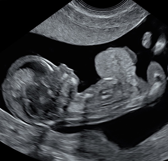
Baby with large exomphalos (abdominal wall defect) at 12 weeks
Structural anomalies: Just Numbers
-
London Pregnancy Clinic specialises in EARLY ANOMALY SCANS:
Fetal Anomalies Explained
Doctors use various terms to describe fetal structural anomalies and related issues:
Baby Structures: Developing fetal organs read our blog.
Structural Fetal Anomalies: General term referring to physical abnormalities in the developing fetus.
Congenital Anomalies: Abnormalities present at birth, including structural defects.
Birth Defects: Conditions that affect the structure or function of one or more parts of the body, present at birth.
Developmental Abnormalities: Irregularities in the growth or formation of organs or body parts during fetal development.
Malformations: Structural abnormalities or irregularities in the formation of organs or tissues.
Dysmorphology: The study of congenital malformations or the abnormal development of an organism.
Teratogenic Effects: Adverse effects on the developing fetus due to exposure to harmful substances during pregnancy, leading to structural anomalies.
Chromosomal Abnormalities: Irregularities in the number or structure of chromosomes, leading to conditions such as Down syndrome.
Genetic Anomalies: Abnormalities in the DNA or genes that can result in structural abnormalities.
Neonatal Anomalies: Abnormalities present in the newborn, often discovered shortly after birth.
Prenatal Screening: Testing and examination conducted during pregnancy to identify potential fetal anomalies.
Prenatal Diagnosis: The process of identifying fetal abnormalities before birth, often through imaging and genetic testing.
Fetal Malformations: Abnormalities in the structure or development of the fetus.
Lethal Fetal Anomalies: Structural abnormalities that are likely to result in fetal or neonatal death.
Doctors often use these words interchangeably in medicine, and how they use them might depend on the specific problem and the doctor or situation.
“Fatal fetal anomaly” means that doctors have found a serious problem with the baby growing inside the mum’s tummy. This problem is so big that it’s very unlikely the baby will survive after birth, or it might not even make it until then. People might also call it a “lethal” or “life-limiting” condition.
It’s a really tough situation for parents because it means their baby is very sick, and it’s unlikely they’ll be able to have a normal and healthy life. Parents might have to make some hard decisions with their doctors about what to do next, like whether to continue with the pregnancy, or if there are other options that might be better for everyone involved.
It’s a sad and difficult time, and the doctors and other healthcare professionals will do their best to help and support the parents through it.
Congenital heart defects (CHD), which are issues with how the heart forms and works after birth, are the most common types of fetal structural anomalies affecting 1 in 100 babies born in the UK.
Among congenital heart defects, the most frequent is called ventricular septal defect (VSD), often referred to as a ‘hole in the heart.’ According to some studies, VSD is the most common condition found in newborns.
The other 10 Most Common Birth Defects include:
- Cleft Lip and Palate: An opening or gap in the upper lip and roof of the mouth.
- Spina Bifida: Incomplete closure of the spinal column, affecting the spinal cord.
- Neural Tube Defects (NTD): Acrania, anencephaly, encephalocele and other anomalies affecting the brain and spinal cord development. Spina bifida is also a type of NTD.
- Gastroschisis: A birth defect where the baby’s intestines protrude outside the abdomen.
- Exomphalos or omphalocele: Birth defect where the baby’s abdominal organs protrude through the belly button area.
- Clubfoot or talipes: Deformity of the foot where it turns inward and downward.
- Limb Reduction Defects: Incomplete or absence of a limb during development.
- Polydactyly: Presence of extra fingers or toes.
- Intestinal and Duodenal atresia: Blockage or absence of a portion of the intestines.
- Kidney Anomalies: Various malformations affecting the development, structure or function of the kidneys.
Additionally, the most frequent minor congenital anomaly in male infants, occurring in 1 in 200 boys, is hypospadias. This condition involves a malformation of the male urethra, resulting in the opening being situated in an atypical position.
Please see below the detailed information about specific fetal anomalies.
Unfortunately, severe structural anomalies are commonly lethal or associated with long-term disabilities. Some of the most severe structural anomalies include:
- Acrania: Absence of the top part of the baby’s skull results in brain dissolution.
- Spina Bifida: A problem where the backbone doesn’t close completely, causing sever issues with the nerves.
- Severe Ventriculomegaly (Hydrocephaly): Too much fluid in the baby’s brain, which can be associated with severe disabilities.
- Hypoplastic Left Heart Syndrome (HLHS): The left side of the baby’s heart doesn’t develop properly, needing complicated surgeries to survive.
- Diaphragmatic Hernia: A hole in the baby’s diaphragm allows organs to move where they shouldn’t, affecting lung growth.
- Exomphalos with Liver: Some organs, including the liver, are outside the baby’s body due to a hole in the belly wall.
- Body Stalk Anomaly: Severe problems with organs in the belly and pelvis, often linked to a short or missing umbilical cord.
- Amniotic Band Syndrome: Fibrous bands in the fluid around the baby can tangle parts, causing deformities or amputations.
- Agenesis (Absence) of Both Kidneys: Both kidneys are missing, leading to life-threatening issues.
- Lower Urinary Tract Obstruction: A blockage in the baby’s urinary tract affects kidney development and can cause lung problems.
- Sirenomelia: The baby’s lower limbs are fused, often with serious problems in the urinary and digestive systems.
- Thanatophoric Dysplasia: A commonest skeletal disorder (dysplasia) causing severe shortening of limbs and a small chest.
- Fetal Hydrops: Abnormal fluid buildup in different areas of the baby’s body, often indicating serious issues or syndromes.
It’s crucial to understand that the seriousness of these issues can differ. The impact depends on factors like the type of problem, any additional complications, and what medical treatments are possible. Every situation is different, so decisions about how to care for and manage these cases require careful thought about each person’s circumstances.
Please see below the detailed information about specific fetal anomalies.
When specific structural anomalies or other physical differences are noticed in the baby during pregnancy, it may suggest a connection to the baby’s genes or chromosomes. In other words, a structural anomaly could be a presentation or symptom of a significant chromosomal or genetic syndrome. These syndromes are collectively known by the broader term “rare diseases.”
Recognizing the link between genes and structural anomalies is crucial for understanding complicated connections between a baby’s genetic code and physical appearance. The same structural abnormality can be an isolated event or represent part of the genetic condition.
Conversely, if it’s determined that the baby has a chromosomal or genetic condition (referred to as a “rare disease”), those structural anomalies serve as physical manifestations of abnormal gene function.
It’s important to note that the prognosis of structural defects associated with chromosomal or genetic conditions is worse than that of isolated anomalies. Many chromosomal and genetic conditions are associated with intellectual disabilities or other significant health challenges.
Soft markers and anatomical variants are synonyms. Other terms used to describe those conditions are chromosomal markers, genetic markers, Down’s syndrome markers, and ultrasound findings of uncertain significance.
Soft markers refer to minor fetal physical features that can be noticed by scan during pregnancy. It’s important to understand that these features are not anomalies or abnormalities but rather subtle variations that might be seen in healthy babies.
They are small details that sonographers and doctors might observe on ultrasound, but they don’t necessarily indicate a problem. These markers are quite common and can be present in perfectly healthy babies. It’s like having unique traits that make each baby special.
If these markers are noticed, your healthcare team will carefully assess and consider various factors to provide you with the most accurate information about your baby’s health. Remember, seeing these markers doesn’t automatically mean there’s a cause for concern.
Soft Markers. Doctors often prefer the more precise term “anatomical variants” or even “ultrasound findings of uncertain significance” when referring to soft markers.
- Absent or Hypoplastic Nasal Bone (NB): The absence or underdevelopment of the nasal bone, which might be a soft marker for certain chromosomal conditions.
- Nuchal Fold (NF): A measurement of the thickness of the skin at the back of the baby’s neck, often considered in prenatal screening.
- Hyperechogenic Bowel: Increased brightness in the baby’s bowel on ultrasound, which might be due to various factors and may resolve on its own.
- Mild (Borderline) Ventriculomegaly: Slight enlargement of the fluid-filled spaces in the brain, which can sometimes be observed in routine ultrasound but may not necessarily indicate a problem.
- Single Umbilical Artery (SUA): Having one instead of the usual two arteries in the umbilical cord, which is often a soft marker but is usually not a major concern.
- Aberrant Right Subclavian Artery (ARSA): An aortic arch branch artery that follows an unusual path, often seen during ultrasound but may not necessarily indicate a problem.
- Intracardiac Hyperechogenic Focus (Golf Ball): A bright spot seen on the baby’s heart during an ultrasound, which is usually harmless but may be associated with chromosomal conditions.
- Mild Dilatation of Kidney Pelvis: Slight enlargement of the kidney’s collecting system, often considered a variant rather than a significant issue.
- Shortening of the Femur or Humerus: Slightly shorter than average thigh or upper arm bones, which may be noticed during an ultrasound but might not pose a problem.
- Choroid Plexus Cysts (CPC): Fluid-filled spaces in the brain, which are often harmless and may disappear on their own, however, they can be associated with Edward’s syndrome (trisomy 18).
- Prenasal Thickness: Thickness of the tissue over the nose, which might be considered as a soft marker during prenatal screening.
- Ductus Venosus (DV) flow: A vessel in the baby’s liver, which might be assessed during certain prenatal screenings.
- Tricuspid Regurgitation (TR): A condition where blood leaks backward through the tricuspid valve of the heart, which can be a soft marker.
- Persistent Right Umbilical Vein (PRUV): The continued presence of a certain vein in the liver is usually a variant but might be considered in prenatal screening.
- Macroglossia: Enlargement of the tongue, which might be observed during ultrasound but could be a normal variation.
- Sandal Gap: Wide spacing between the big toe and the second toe, which might be noted during ultrasound but is typically not a significant concern.
- Rocker Bottom Feet: The shape of the baby’s feet, which may be considered a soft marker in certain situations.
Please see below the detailed information about the most important anatomical variants.
At London Pregnancy Clinic, we specialize in expert ultrasound screenings for fetal anomalies at every scan, adapting our approach based on the stage of gestation. Essentially, every scan from 10 weeks onward serves as an ‘anomaly scan’, although the scope of each scan varies as the pregnancy progresses.
10 Week Scan: A unique offering from our clinic, this advanced 10-week anomaly scan is unparalleled in its early detection capabilities. It aims to identify 10 severe fetal anomalies that are discernible at this stage, making our 10 10-week scan ideal for combining with NIPT.
Early Fetal Scan: Our signature scan, the Early Fetal Scan, involves a comprehensive examination of the developing baby, aiming to detect over 100 serious anomalies. This early detection goes beyond what is typically screened by the NHS at 19-20 weeks, providing reassurance and vital information much sooner.
Anomaly Scan: Our Anomaly Scan, conducted around 22-23 weeks, offers a more extensive review of fetal structures than the standard 19–20-week NHS scan. This thorough examination includes detailed assessments of the baby’s brain, heart, face, fingers, and other vital organs. We also offer additional assessments like uterine artery Doppler and cervical length measurements upon request.
3rd Trimester Anomaly Scan: Recognizing that the NHS does not routinely offer scans after 18-20 weeks, our 3rd Trimester Anomaly Scan fills this gap. Performed at 26-29 weeks, it screens for late-presenting fetal anomalies and monitors fetal growth and well-being. As a comprehensive check becomes technically challenging after 30 weeks, this scan is crucial for detecting late-manifesting anomalies, particularly in the brain, heart, and kidneys, and is an ideal time for clinical 3D/4D imaging.
The London Pregnancy Clinic provides second-opinion scan services for patients with concerns about fetal structural anomalies or anatomical variants.
Using state-of-the-art ultrasound equipment, we specialize in high-resolution fetal imaging at any stage of gestation, starting from 10 weeks.
If you’d like, you can think about having the NIPT done during our second opinion scan. Typically, NIPT isn’t suggested for structural fetal anomalies, but it can be a good option when dealing with soft markers.

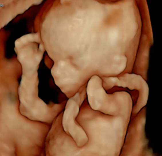
Bay with missing left hand (transverse arm defect): 12 week 3D scan
Common Fetal Anomalies and Anatomical Variants
Achondroplasia
Achondroplasia is a genetic disorder that affects bone growth, and is the most common form of dwarfism. Individuals with achondroplasia typically have short stature, disproportionately short arms and legs, a normal-sized torso, and a characteristic facial appearance. Achondroplasia does not affect intelligence, and individuals with achondroplasia can lead healthy, fulfilling lives. Achondroplasia is not evident during a 20-week scan, but significant bone shortening becomes noticeable in the 3rd trimester. Surprisingly monogenic NIPT, for FGFR3 mutation, can detect achondroplasia as early as 10 weeks.
Acrania
Acrania, a type of neural tube defect (NTD), is a condition where the top part of the baby’s skull, including the brain, does not form properly. This anomaly is often detectable in the first trimester of pregnancy, even at 10 weeks. Unfortunately, acrania is associated with exposed brain tissue and is not compatible with life. While relatively common among structural anomalies, its early detection is crucial for informed medical decisions At the London Pregnancy Clinic, we screen for acrania as early as 10 weeks, prioritising early detection of this severe structural anomaly.
Absence of Cavum Septum Pellucidum
Absence of Cavum Septum Pellucidum (CSP): The absence of cavum septum pellucidum refers to the lack of a small fluid-filled space in the midline of the brain, known as the cavum septum pellucidum (CSP). While some individuals with this condition may not exhibit noticeable symptoms, in some cases, it can be linked to septo-optical dysplasia. It may contribute to problems of vision, developmental issues or other conditions.
Absent Nasal Bone
Absent nasal bone refers to the absence or underdevelopment of the nasal bone during prenatal development. Absent nasal bone does not represent structural anomaly. This anatomical variant is often identified through ultrasound imaging. While the absence of a nasal bone can be a soft marker for certain chromosomal conditions, especially Down’s syndrome, it’s essential to note that it doesn’t necessarily indicate a problem on its own. NIPT can be an excellent solution to reduce anxiety for parents worried about absent nasal bone. Alternatively, they may considered to have diagnostic invasive test.
Agenesis of the Corpus Callosum
Agenesis of the corpus callosum (ACC) is a condition where the bundle of nerve fibers connecting the two hemispheres of the brain, known as the corpus callosum, either partially or completely fails to develop. This anomaly can impact the communication between the brain’s hemispheres. While some individuals with ACC may not show significant symptoms, others might experience developmental delays or intellectual disabilities. The severity of symptoms can vary, making each case unique.
Amniotic Band Syndrome
Amniotic Band Syndrome is a rare condition that occurs during fetal development when fibrous bands from the amniotic sac entangle parts of the fetus, restricting normal development. These bands can lead to various deformities, facial distortion or even amputation of fingers, extremities, or other body parts. The severity of the effects can vary widely. The severe forms of ABS are lethal.
Atrio-ventricular septal defect
Atrio-ventricular septal defect (AVSD) is a common congenital heart defect with a hole in the septum (wall) between the heart’s chambers, affecting all four chambers of the heart (atria and ventricles). This defect can contribute to abnormal blood flow within the heart. AVSD is often associated with Down’s syndrome. The severity of AVSD can vary, and surgical intervention is probably required to correct the defect after birth.
Bilateral renal agenesis
Bilateral renal agenesis occurs when both kidneys fail to develop during embryonic development. This anomaly leads to the absence of both kidneys, essential for filtering waste and regulating fluids in the body. The condition is strongly associated with the absence of amniotic fluid around the fetus, contributing to lung hypoplasia where the lungs may not fully develop. With a practically 100% lethality rate, bilateral renal agenesis is a severe condition.
Body-Stalk Anomaly
Body-stalk anomaly is a rare and severe congenital condition where there is an abnormality in the development of the fetus, leading to the absence of the abdominal wall. This results in the exposure of internal organs outside the body. The baby is also lacking a proper umbilical cord. The condition is associated with severe deformities and is incompatible with life.
Cleft Lip and Palate
Cleft lip and palate are congenital conditions where there is an opening or gap in the upper lip and/or roof of the mouth (palate). These openings occur during embryonic development and can vary in severity. Cleft lip and palate may impact feeding, speech, and facial appearance and can be associated with various chromosomal and genetic abnormalities.
Coarctation of Aorta
Coarctation of the aorta is a common congenital cardiac anomaly where there is a narrowing or constriction in the aorta, the main blood vessel carrying oxygenated blood from the heart to the body. This critical condition requires urgent treatment after birth, often involving surgical intervention to correct the constriction. Generally, CoA may not be easily detectable even by specialist fetal echocardiography.
Congenital Diaphragmatic Hernia
Congenital Diaphragmatic Hernia (CDH) is a congenital anomaly where there is a hole or opening in the diaphragm, the muscle that separates the chest from the abdomen. This opening allows abdominal organs to move into the chest cavity, potentially impacting the development of the lungs. It is crucial to note that diaphragmatic hernia is a severe condition, with a perinatal mortality rate ranging from 30-40%. For survivors, CDH will require surgical intervention after birth.
Duodenal Atresia
Duodenal atresia is a congenital condition characterized by a blockage or closure in the first part of the small intestine, known as the duodenum. This condition is strongly associated with Down’s syndrome. Detectable through prenatal screening, duodenal atresia may create a double-bubble appearance on ultrasound, and it is often linked with polyhydramnios (excess amniotic fluid). The blockage can pose challenges in feeding after birth, necessitating surgical intervention to correct the issue.
Echogenic Bowel
Echogenic bowel refers to an ultrasound finding where the fetal bowel appears brighter than usual. This increased echogenicity can be a soft marker in prenatal screening. It may be associated with various conditions, including intrauterine infections, chromosomal abnormalities (Down syndrome), bowel anomalies, and cystic fibrosis (CF). While often benign, it can also indicate the need for further investigation. It also can be associated with reduced fetal growth.
Exomphalos
Exomphalos (omphalocele) is a congenital condition where the abdominal wall does not fully close, leading to organs such as the intestines, liver, or stomach protruding outside the body and being covered by a transparent membrane. This condition is strongly associated with chromosomal and genetic conditions. Exomphalos requires surgical intervention shortly after birth to place the organs back into the abdomen and close the opening. The surgical outcome will depend on the position of the liver; when the liver is outside the abdomen, some babies may experience problems with lung function.
Fetal Akinesia Deformation Sequence
Fetal Akinesia Deformation Sequence (FADS) is a disorder characterized by reduced fetal movements during pregnancy, leading to various physical deformities. This condition is both clinically and genetically diverse, meaning it can manifest in different ways, and its causes may involve a range of genetic factors. Severe cases of FADS are lethal. Related conditions are Pena-Shokeir syndrome (PSS), arthrogryposis multiplex congenita (AMC), multiple congenital contractures, multiple pterygium and other neuro-muscular disorders.
Fetal Hydrops
Fetal hydrops is a serious condition characterized by an abnormal accumulation of fluid in two or more parts of fetal body, such as the skin, abdomen, and chest. This condition can result from various underlying causes, including heart and chromosomal or genetic abnormalities. Fetal hydrops is associated with a significant risk to the health of the developing fetus and may lead to severe complications and death.
Gastroschisis
Gastroschisis is a congenital condition where an opening in the abdominal wall allows intestines to protrude outside the body. Free-floating intestines in gastroschisis may be susceptible to damage from mechanical friction and amniotic fluid toxicity. Gastroschisis requires surgical intervention shortly after birth to place the organs back into the abdomen and close the opening. The general outcome of surgery is favorable in the majority of cases. Complicated cases may involve liver herniation or result in the loss of part of the intestine.
Holoprosencephaly
Holoprosencephaly (HPE) is severe embryonic brain malformation. It is characterized by the incomplete separation of the embryonic brain into distinct hemispheres. Holoprosencephaly can lead to intellectual and developmental disabilities and facial deformities. The severity of the condition can vary widely, however majority of individuals with HPE experience profound disabilities or die. At the London Pregnancy Clinic, we screen for holoprosencephaly as early as 10 weeks, prioritising early detection of this severe brain anomaly.
Hypoplastic Left Heart Syndrome
Hypoplastic Left Heart Syndrome is the most severe common congenital heart condition where the left side of the heart, including the left ventricle and aorta, is underdeveloped. This complex anomaly hinders the heart’s ability to pump oxygenated blood effectively. HLHS is a critical condition requiring urgent medical intervention, and after involving a series of surgeries. It is also associated with intellectual delays in affected babies. Due to the absence of the left part of the heart, creating a normal four-chamber heart, even with advanced cardiac surgery, is impossible.
Limb reduction defects
Limb reduction defects are congenital anomalies characterized by the underdevelopment or absence of limbs during fetal development. These defects can affect the arms, legs, or both and vary in severity, and the causes can be genetic or environmental. Statistics from the National Congenital Anomaly and Rare Disease Registration Service in England reveal that limb defects are among the most common congenital abnormalities and, paradoxically, less detectable during prenatal ultrasound screening.
Megacystis
Megacystis is a prenatal condition characterized by an abnormally enlarged bladder (more than 7 mm in length) in the developing fetus, typically observed around 11-13 weeks. This condition can be associated with underlying structural or functional abnormalities of the urinary system, with the worst-case scenario being the development of lower urinary tract obstruction (LUTO). While isolated small megacystis may resolve on its own, it can also signal more complex issues, including chromosomal abnormalities.
Polydactyly
Polydactyly is a congenital condition characterized by the presence of extra fingers or toes. This anomaly can range from a small, non-functional digit to a fully formed and functional extra digit. Polydactyly is often isolated, however, it can be associated with chromosomal abnormalities, such as trisomy 13 (Patau syndrome), and many different genetic syndromes.
Renal pelvic dilatation (RPD)
Fetal renal pelvic dilatation (RPD) occurs when there’s swelling or enlargement in one or both of the kidneys. In more severe cases, this enlargement is also known as hydronephrosis. The swelling happens because urine is not draining properly from the kidney to the bladder or down from the bladder. This can be caused by various factors, such as a blockage in the urinary tract or a condition affecting the valves that control the flow of urine. In cases of severe blockage, the baby may suffer from a lack of amniotic fluid. It’s crucial to monitor and, if necessary, provide medical intervention after birth to ensure proper kidney function.
Right Aortic Arch
A right aortic arch (RAA) is an anatomical variant in the aorta’s structure, characterized by its position on the right side instead of the usual left. While generally asymptomatic, RAA can exert pressure on the trachea (windpipe) and oesophagus (pathway from mouth to stomach), affecting breathing and swallowing. Evaluation through detailed imaging, especially fetal echocardiography, and a comprehensive head-to-toe assessment of anatomy and associated conditions is crucial. Moreover, there’s a small chance of an association with 22qdel syndrome, a genetic disorder called DiGeordge syndrome.
Talipes
Clubfoot, also known as talipes, is a common congenital condition where a baby’s foot is twisted out of shape or position. This condition can affect one or both feet. The foot may be turned downward and inward, making normal movement difficult. Clubfoot is usually treatable with early intervention, including casting, bracing, and, in some cases, surgery. In rare cases, talipes can be an early presentation of Fetal Akinesia Deformation Sequence (FADS) or other musculoskeletal disorders.
Tetralogy of Fallot
Tetralogy of Fallot (TOF) is a common congenital heart condition. Before birth, TOF consisted of three abnormalities: a VSD (hole in the heart’s wall), narrowing of the pulmonary valve and overriding aorta (aorta positioned over the VSD). This complex condition can lead to oxygen-poor blood being pumped into the body. Tetralogy of Fallot is associated with various chromosomal and genetic conditions and structural anomalies. TOF will require surgical intervention after birth.
Thanatophoric dysplasia
Thanatophoric dysplasia is severe skeletal dysplasia characterized by abnormal bone development, leading to a small ribcage, short limbs, and a disproportionately large head. The term “thanatophoric” is Greek for “death-bearing,” highlighting the severe lethal nature of the condition. Being rare, TD is the most common lethal skeletal disorder and can be diagnosed by monogenic NIPT for FGFR3 mutation.
Transposition of the Great Arteries
Transposition of the Great Arteries (TGA) is a common congenital heart condition where the main arteries, the aorta and the pulmonary artery, are wrongly switched. This leads to an abnormal circulation of oxygenated and deoxygenated blood. TGA is a critical condition requiring urgent medical intervention immediately after birth. It is a congenital anomaly where prenatal detection makes an enormous difference in a baby’s life. Undetected TGA left without urgent postnatal treatment can result in neonatal death.
Ventricular Septal Defect
A Ventricular Septal Defect (VSD) is a prevalent heart anomaly characterized by a hole in the wall that separates the heart’s lower chambers (ventricles). This results in a passage for blood between the ventricles, essentially creating a “hole in the heart.” The opening size can vary, and while some VSDs may close naturally over time, others might require surgical intervention. VSD often occurs as part of more complex heart anomalies. Fetal echocardiography is essential if there are any suspicions of a VSD, providing detailed insights into the heart’s structure.
Ventriculomegaly
Ventriculomegaly is the enlargement of the brain’s fluid-filled spaces, known as the ventricles. This condition is often identified through ultrasound imaging around or after 20 weeks of pregnancy. Ventriculomegaly is the most common fetal brain abnormality and can be the manifestation of other more severe neurological conditions. While mild cases are benign and may not lead to any issues, severe ventriculomegaly (hydrocephaly) can be associated with neurological abnormalities and intellectual disabilities. Close monitoring and additional diagnostic tests (neurosonography, MRI, infection screen, amniocentesis) may be recommended to assess the overall health and development of the baby.
No, certainly not. The list only covers common structural and cardiac anomalies usually detectable by scans. There are hundreds of other congenital defects not included in the list. For instance, severe eye anomalies that can lead to blindness or complex genito-urinary abnormalities like a cloacal anomaly.
Yes, rare structural anomalies can be identified through ultrasound scans, although their detection may pose challenges due to their infrequency and complexity. Routine ultrasound scans, like 20-week anomaly scans, primarily concentrate on common structural anomalies, and further, specialized scans or diagnostic tests may be necessary for a more thorough assessment.
Some of the rare anomalies are better detectable in the 1st trimester. At London Pregnancy Clinic we specialise in expert ultrasound for early detection of fetal anomalies from 10 weeks. We also provide second-opinion scans for rare fetal anomalies.
Our clinic strongly emphasises early diagnosis of fetal anomalies because identifying potential health concerns at an early stage allows for a more thorough understanding and effective management. Early detection enables healthcare professionals to provide detailed information about the anomaly, its potential impact, and the available options for care and intervention. This proactive approach empowers parents to make informed decisions, facilitates appropriate medical planning, and ensures that necessary support and resources are in place.
It’s essential to note that many advanced genetic tests, such as microarrays and exome sequencing, as well as extended non-invasive prenatal testing (NIPT) options, may require several weeks to deliver results. Therefore, an early diagnosis ensures that there is sufficient time for these comprehensive tests and allows for a more comprehensive and thoughtful approach to the overall care and support provided during the pregnancy journey.
We are truly sorry to hear that you’ve received such challenging news. Dealing with a severe fetal anomaly can be incredibly difficult.
The London Pregnancy Clinic provides specialized second-opinion scans for those contemplating a second opinion. Our focus is on delivering thorough and comprehensive fetal assessments, offering valuable insights that can aid in confirming the diagnosis and providing additional information about your baby’s condition.
We understand that waiting for the scan adds to the stress and uncertainty, and we are committed to arranging your appointment as promptly as possible.
Feel free to get in touch with us through:
- Email: info@londonpregnancy.com
- Phone: 020 3687 2939
- Or, conveniently book your appointment through our online booking system.
We are here to support you during this challenging time.
Determining the optimal time for the anomaly scan is somewhat nuanced. Technically, the most suitable period is around 22-23 weeks when the baby’s structures are more developed, enhancing visibility on the scan. Importantly, at this stage, critical brain structures such as the corpus callosum and vermis have formed. This timing is also preferable for 3D evaluation of the face, especially in cases requiring medical 3D imaging.
However, there’s a strategic consideration. If an unexpected anomaly, particularly associated with chromosomal or genetic conditions, arises, conducting the scan at 22-23 weeks may pose challenges due to the delayed results of genetic tests (via amniocentesis). In such scenarios, opting for the scan at 19-20 weeks allows more flexibility while awaiting results.
In practical terms, if you’ve received normal results from Non-Invasive Prenatal Testing (NIPT) and an early fetal scan (early anomaly scan) was normal, you may choose to delay the anomaly scan to 22 weeks.
Opting for this timing enhances the likelihood that the information about your baby’s condition will be more accurate and precise. It’s important to note that this recommendation is specific to our or other non-NHS services, as the timing for the anomaly scan in NHS is regulated by strict guidelines.
The National Congenital Anomaly and Rare Disease Registration Service (NCARDRS) in England, operated by the government through Public Health England, is a comprehensive registration service. It systematically gathers and ensures data quality related to congenital anomalies and rare diseases in England.
Currently, NCARDRS collects data on over 1,400 different congenital anomalies and rare diseases and has achieved 100% coverage of births in England since 2018.
Yes, there is some inconsistency in information regarding the NHS Fetal Anomaly Screening Program (FASP) screening for only 11 anomalies, while the National Congenital Anomaly and Rare Disease Registration Service (NCARDRS) collects information about over 1400 different congenital anomalies and rare diseases for babies born in England.
In the NHS, sonographers typically perform a routine anomaly scan that focuses on the detection of major structural anomalies. While the primary focus is on identifying the 11 standard anomalies outlined in the fetal anomaly screening program, the scan also involves a comprehensive assessment of the baby’s overall development and well-being. Sonographers will thoroughly examine various structures and organs to identify any potential abnormalities.
It’s important to note that sonographers in the NHS perform an excellent job, extending their assessments to detect a broader range of anomalies through ultrasound. The list of 11 anomalies outlined in NHS guidelines may primarily be for audits and quality assurance purposes.
For your peace of mind, it may be helpful to have a chat with sonographers to better understand the details of the anomaly scan they carry out. This way, you can gain a clearer picture of the scope and purpose of the 20-week anomaly scan examination.


