
Welcome and brief history
A sonographer greets you and checks your details and how many weeks pregnant you are. No special preparation is needed for a late-pregnancy scan.
Checking the position of your baby to exclude breech presentation and fetal wellbeing
Usually performed from around 36 weeks, this late-pregnancy ultrasound confirms whether your baby is head down, breech or lying sideways, and helps you and your maternity team plan the safest birth.
Available atLPC City (E1)LPC West (SW10)
The main aim of the Presentation Scan is to diagnose breech position or other abnormalities in the baby’s position. The clinician assisting you with the delivery will use this information to prepare for your baby’s birth!
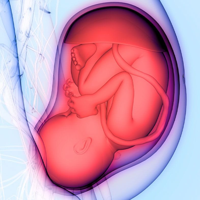
"Presentation" describes which part of your baby lies closest to the birth canal, while "lie" describes the way your baby's body is angled in the womb. By the final weeks of pregnancy most babies settle head down, known as cephalic presentation, which is the most common position for a straightforward vaginal birth. Towards the end of pregnancy a small number of babies remain bottom or feet first, a position called breech (RCOG).
Breech simply means your baby is bottom or feet first. There are three main types, and an ultrasound can tell them apart (Tommy's):
Less commonly, a baby may lie sideways, called a transverse lie, or at an angle, called an oblique lie. Because a sideways or unstable lie in the last weeks affects how and where your birth is planned, confirming exactly how your baby is lying matters — and that is what a presentation scan is designed to do.
A few general figures that put late-pregnancy positioning in context. They describe the wider population, not a prediction for any one pregnancy.
The main aim of the Presentation Scan (or Pre Delivery Scan) is to diagnose breech position or other abnormalities in the baby’s position. We also perform thorough checkup of fetal growth and estimate fetal weight. We assess fetal wellbeing by umbilical artery and middle cerebral artery Dopplers.
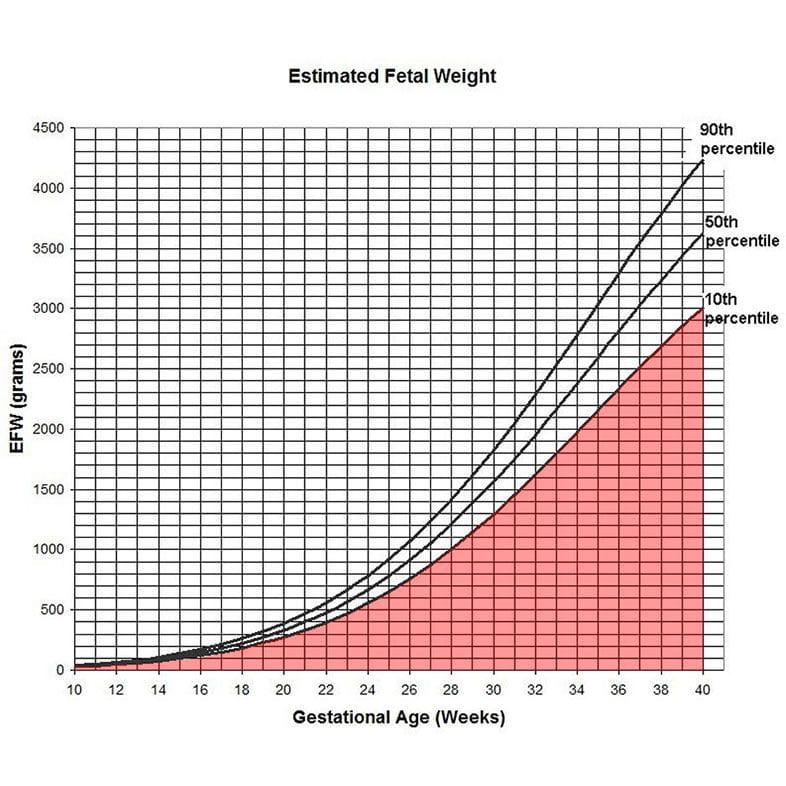
Normally, a few weeks prior to birth, the baby will move into a delivery position where the head is positioned close to the birthing canal. However, about 3-4% of babies remain in a breech position (RCOG). This is where the baby's buttocks are positioned closest to the birthing canal, resembling a sitting position.
Delivering a baby in breech position can be dangerous, but in roughly 50% of cases (RCOG) the position can be corrected by external cephalic version (ECV), which is a manual process performed by an obstetrician to turn the baby upside down in the womb allowing a head-first delivery. Alternatively, a caesarean section may be the safest option.
A presentation scan is usually arranged in the final weeks of pregnancy, from around 36 weeks. You may find it reassuring if any of the following apply.
You would like to confirm whether your baby is head down before finalising your birth plan
Your midwife has felt that your baby may be breech or lying sideways during an abdominal examination
You are considering an external cephalic version (ECV) and want your baby's position confirmed first
You have had a breech baby or a breech birth before
You would simply value peace of mind that all is on track as your due date approaches
In most cases, an experienced midwife can recognise a breech position through an external manual examination of the baby, which is done by feeling for the baby’s orientation through the mother’s lower tummy. However, in some cases, the breech position remains unnoticed until active labour and/or rupture of membranes.
Thus, the main aim of our Baby Position Scan is to recognise an undiagnosed breech position.
Some of the other things we also look out for include:
A sonographer greets you and checks your details and how many weeks pregnant you are. No special preparation is needed for a late-pregnancy scan.
Warm gel is applied to your bump and the sonographer gently moves a probe over your abdomen to see how your baby is lying: head down, bottom first or sideways.
We estimate your baby's weight and look at the amniotic fluid, the position of the placenta and blood flow in the umbilical cord using Doppler measurements.
A specialist fetal medicine clinician reviews the images and talks you through what they mean for your birth plan, including whether your baby is head down.
You leave with a clear summary of the findings to keep and to share with your midwife or obstetric team.
What we check
If your baby is head down (cephalic), that is the most common position for the final weeks and is generally reassuring for a planned vaginal birth. Your report confirms this alongside your baby's estimated weight, the amniotic fluid and the position of the placenta.
If your baby is breech or lying sideways, knowing this before your due date gives you and your maternity team time to plan. Once breech is confirmed at or after 36 weeks, national guidance recommends discussing the benefits and risks of the options available — an external cephalic version (ECV) to try to turn your baby head down, a planned caesarean birth, or a vaginal breech birth (NICE). These decisions are always made with the NHS or private maternity team looking after you.
Our role is diagnostic: a presentation scan gives you clear, up-to-date information, but it does not replace your antenatal care. If you would value a fuller third-trimester assessment, our growth scan looks in detail at your baby's size over time, our wellbeing scan combines growth and wellbeing checks in one appointment, and our third trimester anomaly scan looks for the small number of conditions that can develop late in pregnancy. For current prices across our scans, see private pregnancy scan costs.
If you ever notice a change in your baby's movements or have any concern about your baby's wellbeing, contact your midwife or maternity unit straight away rather than waiting for a scan.
The best time for this scan is at 36 weeks of pregnancy.
By this time, if the baby’s head is not near the birthing canal, there is still time to discuss external cephalic version (ECV) or other delivery plans with your doctor or midwife.
The Baby Position Scan is less effective at detecting anomalies for the following reasons:
Usually performing a 4D scan during a Baby Position scan is ineffective because:
We are happy to provide you the images of your baby, however in majority of the cases it will be impossible to obtain them. Babies at the end of the pregnancy usually facing backwards and it is not feasible to get any images.
Advice on the COVID-19 vaccine has changed since the pandemic. Pregnancy on its own no longer makes you eligible for an NHS COVID-19 vaccine: the NHS now offers it to people aged 75 and over, residents of care homes for older adults and anyone with a weakened immune system. If you are in one of those groups, you can still have it while pregnant or breastfeeding. Check the NHS website or ask your midwife or GP whether you are eligible.
The vaccines every pregnant woman is offered free on the NHS are the flu jab (in flu season, at any stage of pregnancy), whooping cough (from 16 weeks, ideally before 32 weeks) and RSV (from 28 weeks). Our vaccines in pregnancy guide explains the timing of each.
A presentation scan — also called a pre-delivery or baby position scan — is an ultrasound in the final weeks of pregnancy that checks which way your baby is lying: head down (cephalic), bottom or feet first (breech), or sideways (transverse). It also reviews your baby's estimated weight, the amniotic fluid, the position of the placenta and blood flow through the cord, giving you and your maternity team a clear picture before birth.
Most babies settle head down by around 36 to 37 weeks, and once there is less room it becomes less usual for a baby to turn on its own (Tommy's). It can still happen. If your baby stays breech, your team may offer an external cephalic version (ECV), which turns around half of breech babies (RCOG). A presentation scan confirms exactly how your baby is lying so these options can be discussed with your midwife or obstetrician.
The two overlap but have different emphases. A presentation scan focuses on how your baby is lying in the final weeks so you can plan your birth, while a growth scan concentrates on measuring your baby's size and tracking growth over time. Both also check the amniotic fluid, placenta and blood flow. If you would like a fuller third-trimester check, our wellbeing scan combines growth and wellbeing in one appointment.
National guidance recommends that if a breech position is suspected on abdominal examination at or after 36 weeks, an ultrasound scan is offered to confirm how your baby is lying (NICE). Feeling your bump from the outside is helpful but not always conclusive, so a scan removes the uncertainty and shows precisely which way your baby is lying.
Yes. Ultrasound has been used in pregnancy for many years and is considered safe when carried out by trained professionals. The scan is external and painless, and it does not use any radiation. If you have any concern about your baby's wellbeing, contact your midwife or maternity unit rather than waiting for a scan.
Most babies are breech at some point in pregnancy and turn head-down on their own before term. If your baby is still breech late on, the scan confirms the position and the type of breech, and your team will talk through the options with you.
Our breech presentation page explains how often breech resolves by itself, what ECV involves and how likely it is to work, and what the evidence says about the birth options.
No. The flu vaccine is recommended at any stage of pregnancy, right up until you go into labour. The whooping cough and RSV vaccines can also still be given late in pregnancy — the earlier windows (from 16 and 28 weeks respectively) are simply the ideal, because your antibodies need time to build and cross the placenta before birth. All three are free on the NHS.
Our flu, whooping cough and RSV vaccine guides explain what late vaccination does and does not change.
Contact
The figures and clinical statements on this page are drawn from the sources below. Guidance evolves — always discuss your individual circumstances with a clinician.
Cookies on this site
We use cookies needed to run this site. With your permission we would also use analytics and advertising cookies to see how the site is used and which of our adverts lead to bookings. Declining will not affect your care or your booking. See our Privacy notice and Terms & Conditions.