
Tag: birth

How big is the Embryo at 8 weeks?

Most pregnancy apps will make you aware of the size comparison of your baby against a fruit/ vegetable/ animal or even baked goods, but rarely do you see images showing just how your baby would fit into the palm of your hand!

Take a look at this image which shows you the real size of your baby at 8 weeks!

At 8 weeks Dr Ushakov will be able to check for the heartbeat, date the pregnancy, check for single/multiple pregnancies. He will also check the structures of the pregnancy at this stage including the gestational sac, yolk sac and placenta.

During the Viability scan you will find out the estimated due date (Which will give you plenty of time to pack up that hospital bag and plan out your babymoon)!

If you are interested in having the viability scan at City Ultrasound, you can book our scans or give us a call on 02036872939 for more information

Choosing Your Scan

Choosing Your Scan ?

It can feel a difficult decision knowing when to book in for your ultrasound scans, but truth be told there is no right or wrong time. Whether you are seeking general reassurance, a comprehensive check of your baby’s structures or an NIPT we have scans to suit your individual needs. Your baby is developing at such a pace and week by week there is visible growth. We offer scans for each and every stage of your baby’s development so you can feel assured at every step of the way.
For the best diagnostic performance, we have created a comprehensive breakdown of our scanning options and the prime time to have them done:

?6-9 weeks – Viability Scan

?10-16 weeks – Early Fetal Scan

?20-23 weeks – Anomaly Scan

?28- 34 weeks – Baby development Scan

?36-40 weeks – Baby Position Scan

?8-40 weeks – Scan Upon Request

To find out more details regarding the above scans please check out our website which has a breakdown of everything included in each scan.

Fred Ushakov – Teaching

One of Dr Fred Ushakov’s passions is teaching and training other professionals in the field of Fetal Medicine and the applications of ultrasound screening. Prior to the pandemic Fred lectured in countries such as the US, Australia, Italy, Sweden, Peru, Egypt, Romania, Ukraine and China, where the picture below was taken. Fred has also collaborated with GE Healthcare in live masterclass demonstrations in their Brussels HQ.

During the pandemic, Dr Ushakov was one of the pioneers to start broadcasting his lectures online. His lectures are mainly focused around his field of interest of early anomaly detection and the Early Fetal Scan. In the past year alone, over a thousand ultrasound professionals have attended his courses online.

Folic Acid

What is folic acid? 

Folic acid is a synthetic (man-made) form of the Vitamin B-9 (folate), which naturally occurs in some foods such as asparagus, peas and broccoli. Folate supports generation of healthy new red blood cells, which carry oxygen around the body.  The deficiency of red blood cells may be conditions like anaemia, which often causes weakness and fatigue.  

Why is folic acid often recommended to women during their pregnancies? 

Prior to conception and within the first trimester (around week 12) it is often recommended that women take this artificial supplement to reduce the risk of a neural tube defects and ensure the healthy formation of red blood cells. Taking folic acid Folic acid also supports maternal tissue growth and increases maternal folate to reduce risk of neural tube defect. Can reduce the risk of developing neural tube defects by up to 70% (WHO, 2015).  

What is a neural tube defect? 

The neural tube is an embryonic structure that lays the foundation for the central nervous system (consisting of the brain and spinal cord). Neural tube formation begins just three weeks after conception, and proper formation is vital for brain development. Neural tube defects occur when the formation of the neural tube does not develop or form correctly. These may result from a lack of folic acid in the first 28 days of pregnancy, causing the neural tube to poorly develop. Typically, the neural tube closes in the 4th week following conception. Neural tube defects include birth defects of the skull and brain like acrania or encephalocele, spine and spinal cord, such as spina bifida.  

How much folic acid is recommended? 

It is recommended that those who are planning to get pregnant take 400 micrograms supplement daily (NICE, 2014) both prior to pregnancy and for the first 12 weeks. The NICE recommend taking this supplement regardless of whether you are eating folate-rich foods.   

Your GP may prescribe or recommend a higher dose if you have a BMI over 30, have diabetes, anaemia, sickle–cell disease, coeliac disease, take medication for epilepsy or have a history of neural tube defects.  

What foods contain folic acid? 

Asparagus, peas, broccoli, oranges, bananas. Green beans, kale, spinach, yeast extract and fortified cereals.  

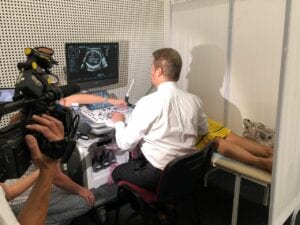
Can you detect neural tube defects by ultrasound?

Yes – Dr Ushakov has been developing new methods for detecting these anomalies in the first trimester from as early as 10 weeks.  

For support for spina bifida and hydrocephaly: Shine Charity 

Sources

NICE (2014). Maternal and Child Nutrition [Online] [link

NICE (2021). Neural Tube Defects (Prevention in Pregnancy) [Online] [link

SHINE (2021). Folic Acid. [Online] [Link

Ushakov, F., Sacco, A., Andreeva, E., Tudorache, S., Everett, T., David, A.L & Pandya, P. P. (2019). Crash Sign: New First‐Trimester Sonographic Marker of Spina Bifida. [Online] [Link

World Health Organisation (2015). Prevention of Neural Tube Defects by The Fortification of Flour with Folic Acid: A Population-Based Retrospective Study in Brazil. [Online] [link]

Fetal and Maternal Heart Rates

Did you know that your baby’s hear rate during pregnancy is in the range of 110-160 beats per minute? Your baby’s heart is working extra hard while it is growing and developing. The mother’s heart rate also increases during pregnancy as the amount of blood it pumps increases by 30-50% during gestation and can average at 70-90 beats per minute.

Studies have shown that the average heart rate rises steadily through pregnancy. At 10 weeks GA, the average heart rate is ~79 BPM. By 40 weeks GA, the average rate was ~89 BPM.

In today’s video you can see a 2D video of the so-called 4 chamber view (4CV) of the fetal heart at about 14 weeks. Ultrasound is an important tool for the screening of Congenital Heart Defect (CHD) which is the most common type of anomalies that could occur during pregnancy. According to the NHS website: “Congenital heart disease is one of the most common types of birth defect, affecting up to 8 in every 1,000 babies born in the UK.”

At City Ultrasound our approach is to monitor the fetal heart function and development at every scan we do. Echocardiography is the examination of the fetal heart using ultrasound and is included with every scan where possible.

Tricefy – Secure Cloud for your Baby Images

We know that sharing your pregnancy scan experience is important, especially in light of the current pandemic restrictions. That’s why we’ve partnered with triceimaging to be able to send you the images, videos and scan report straight to your mobile phone (as well as giving you some printouts…)

From @triceimaging: Don’t have a belly to show off yet? Ultrasound pictures will do!

With a push of a button – The Tricefy Patient App allows you to send patient images directly to your loved ones. Click on the link in our bio to refer your provider (and try our demo?!) and we will set them up with a free trial so that they can start sending quick, secure, high-resolution links of your ultrasound scans directly to your mobile phone!

Hosting BBC at City Ultrasound

Featuring on the BBC ??

We are extremely honoured to have been approached by the BBC to take some stock images and videos for their archive! The BBC producers came across some of our ultrasound images and were ‘really impressed by the outstanding quality and clarity of the images’.

We enjoyed hosting the BBC producers and camera crew at our clinic and talking about the importance of prenatal ultrasound screening and especially Dr Ushakov’s areas of expertise in echocardiography and neurosonography.

This means that going forward you will see our images on BBC news on items related to fetal medicine and pregnancy. We would like to clarify that, to date, City Ultrasound is not related to any of those news items.

Ultrasound Gel

What is it and why do we need it? As you may remember from our post about how ultrasound works, you’ll recall that ultrasound tech utilises sound waves.

Those waves travel through the mother’s tummy and are reflected to the transducer when they reach the surface the clinician would like to examine. Ultrasound waves struggle to travel through air, so when transabdominal ultrasound is performed, we apply a gel to remove any interference from air particles in between the mother’s tummy and the transducer.

This is also the reason why in older ultrasound equipment a full bladder is required to enhance the imaging quality – this is not the case with our advanced Voluson E10!

What are the ingredients you ask? Mainly water and propylene glycol, which is substance often used in food, cosmetics and hygiene products. It is designed to be sticky to allow it to be spread on the tummy without running off.

At City Ultrasound we use a purposely designed ultrasound gel warmer to avoid the unpleasant sensation when a cold gel is applied. Because the small details matter…

Baby Boy or Baby Girl?

Is it a boy or is it a girl? ♂️♀️???

baby boy example

Can you guess by looking at the above ultrasound image? The gender of the baby can be determined using ultrasound from about 13 weeks of gestation with a high certainty of about 95%. From 14 weeks, the gender can be determined with close to 100% certainty using ultrasound.

Another way the sex of the baby can be determined is through Non-Invasive Prenatal Testing (NIPT) from 10 weeks of gestation – however results typically take 5-7 days to come back from the lab.

At City Ultrasound, we can determine the gender of your baby from 13 weeks upon request. This is included organically in the cost of the scan, however the primary focus of our specialist would be to check the wellbeing and development of your baby. We don’t offer a quick ‘gender scan’ just to determine the sex of the baby

On our website, we have in-depth information about our process and types of scans, but if you do have any questions at all, please feel free to get in touch with us!

✅ Call us on 020 3687 2939

✅ Email us on info@cityultrasound.co.uk 

Birthdays

In today’s post we’s like to deviate a bit from the usual ultrasound pregnancy scan content to talk about birthdays! As the world is celebrating the birth of 2021 we decided to look at some stats on the ONS website. Ever wondered how common is your birthday?

We looked at some stats from the ONS and about average daily births in England and Wales from 1995 to 2014. It’s not a big surprise that the least common birthdays coincide with national holidays: 26th Dec, 25th Dec and 1st Jan are the least popular birthdays according to ONS data (in this order.)

The curious thing is that the most popular birthday period comes towards the end of September, and in particular September 26th being the most common birthday (followed by September 25th and 24th.) September 26th is 39 weeks and 2 days after Christmas, which is approximately 9 months… Hmmmm, looks like a lot of babies are being made right now during the festive season!

City Ultrasound was born on 12th of April which is the 305th out of 366 in the list. How popular is your birthday? Visit the ONS website to find out!

Source: ONS website. Note that February 29th is actually the least common birthday, but since it only occurs every 4 years the data was adjusted to account for that.

  • 1
  • 2