Week 4 of Pregnancy

Week 4 of your Pregnancy
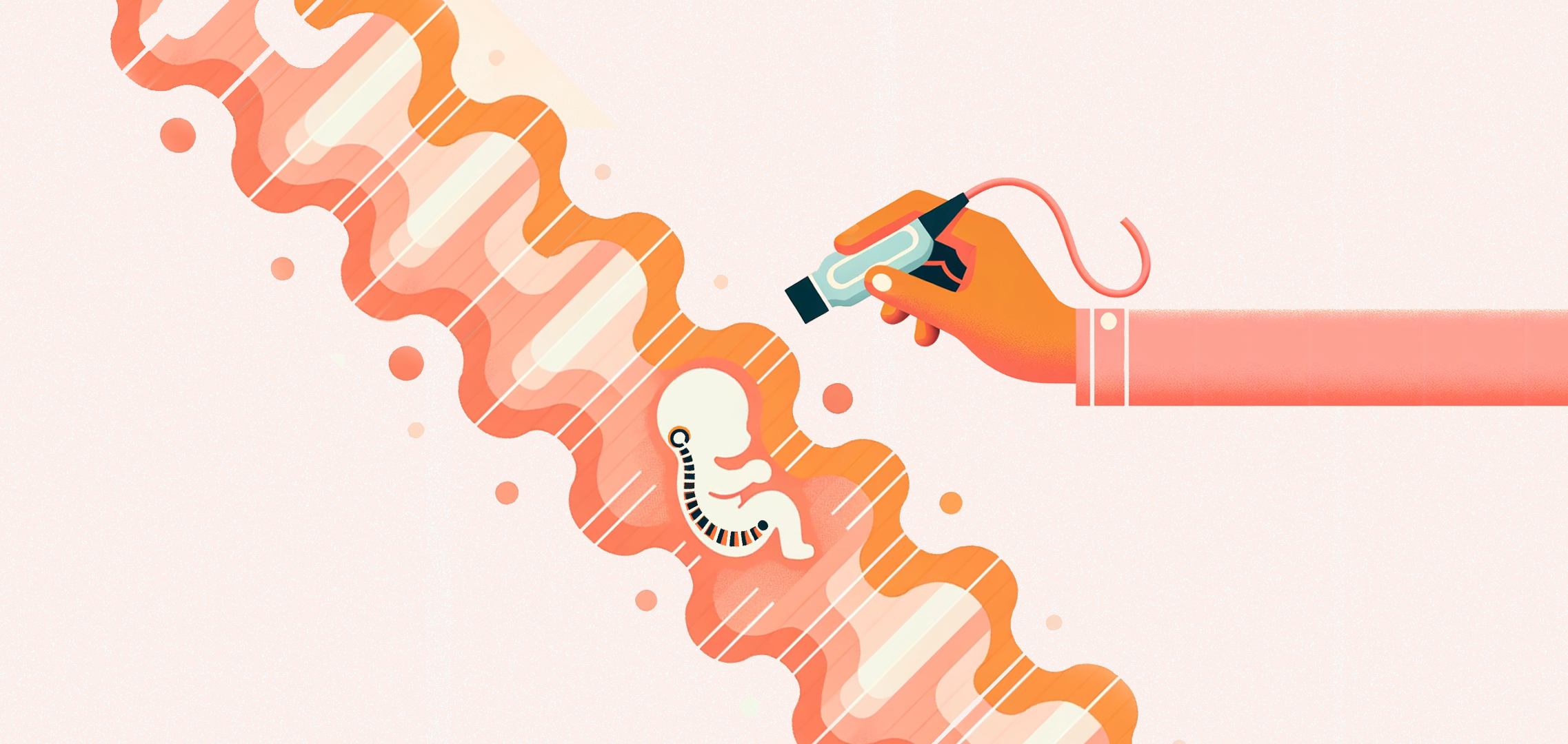
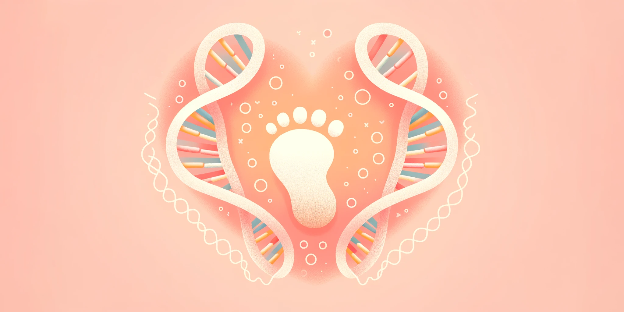
Your Baby’s Remarkable Beginning
-
Published
-
Last Modified
Tags
This blog post is part of a series that breaks down early pregnancy week by week. Today we will be talking about Week 4 of your pregnancy journey! Within this article, we’ll delve into the astonishing advancements occurring during this initial phase of pregnancy. Although your baby remains incredibly minuscule, measuring at no more than 1 millimeter (similar to a poppy seed), a realm of growth and transformation is already well underway.
Key Concepts: Gestational Weeks vs. Post-Conception Weeks
Understanding the difference between gestational weeks and post-conception weeks is crucial for accurately tracking the progress of your pregnancy.
Gestational Weeks: Gestational age refers to the age of the pregnancy and is measured from the first day of the mother’s last menstrual period (LMP). This is the standard method used by healthcare providers to track pregnancy. For example, at 4 weeks gestation, the embryo is about 2 weeks post-conception. Gestational age is crucial for determining the due date and monitoring the baby’s development.
Post-Conception Weeks: Post-conception age, also known as embryonic age, is measured from the time of conception. It provides a more precise timeline of the baby’s development. For instance, at 4 weeks post-conception, the embryo is undergoing significant developments, such as the formation of the placenta and the beginning of the amniotic sac.
Pregnancy Checklist at 4 Weeks
While it’s still early in your pregnancy, there are some important steps you can take:
- During the initial 4 weeks of pregnancy, you may not experience noticeable symptoms. To verify your pregnancy, consider taking a pregnancy test.
- Once you’ve confirmed your pregnancy you may think about your first prenatal visit with your doctor, even though it may not occur for another couple of weeks. With us, this would be our Viability/Dating Scan. During this visit, our specialists, like Miss Shaz Khojasteh, will check for single/multiple pregnancies, date the pregnancy and check the main structures of the gestational sac. We believe early pregnancy scans are essential for peace of mind and early reassurance for parents.
- Focus on adopting healthy diet and lifestyle habits. Say goodbye to smoking and alcohol, opt for nutritious foods, and stay well-hydrated.
- The NHS provides valuable recommendations for expectant mothers, including considering the importance of taking supplements during pregnancy. Additionally, it’s essential to recognise how emotional changes that often accompany pregnancy can impact your relationships.
Developmental Milestones: Week 4
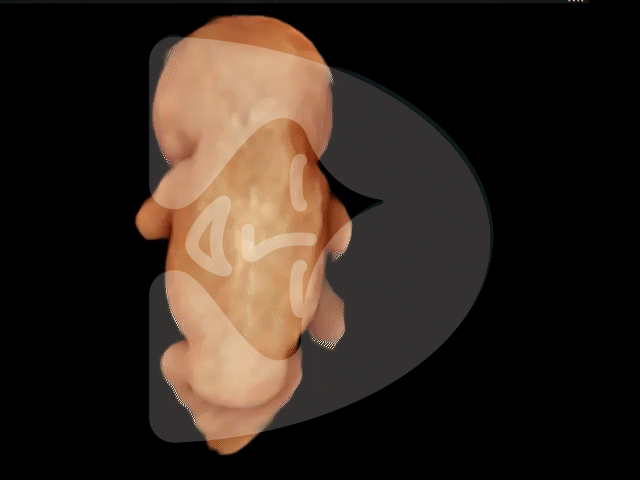
By Week 4, your baby develops from being a single-cell zygote to a blastocyst. Rapid cell division shapes the future. At this stage, your embryo completes its journey from the fallopian tube to the uterus, burrowing into the uterine lining. Half becomes your future child, and the other forms the placenta—a vital nutrient carrier.
The amniotic sac, often called the “bag of waters,” forms around the embryo, along with the yolk sac, important for your baby’s digestive system.
Your embryo now consists of three unique cell layers:
- The endoderm—nurturing your baby’s digestive system, liver, and lungs.
- The mesoderm—laying the foundation for your baby’s heart, sex organs, bones, kidneys, and muscles.
- The ectoderm—shaping your baby’s nervous system, hair, eyes, and outer skin layer.
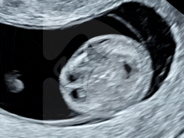
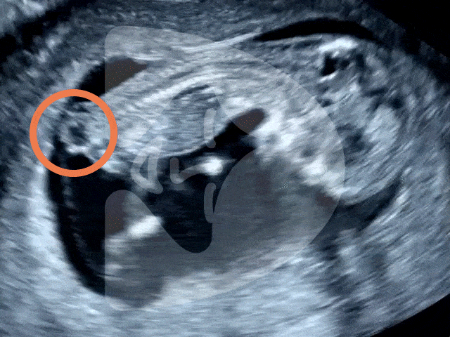
What is seen on the Ultrasound: Week 4?
One of the common queries we receive at the London Pregnancy Clinic is whether having an ultrasound at 4 weeks gestation is a requirement. At this stage, around the fourth week of pregnancy, significant developments are occurring. The blastocyst is in the process of dividing into an embryo and placenta. However, it’s crucial to understand that an ultrasound of your uterus during this early stage will typically reveal what appears to be a minuscule dot known as the gestational sac, and it’s important to note that a heartbeat is not typically detectable at this early point of development. Detecting a more advanced pregnancy may require waiting until a later stage.
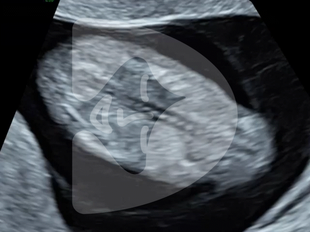
Thinking ahead: Week 5?
The next steps in your pregnancy journey involve preparing for your first ultrasound appointment. It’s completely normal to eagerly anticipate your first ultrasound, but it’s essential to be aware that at 4 and 5 weeks of gestation, the gestational sac is typically too small to be easily visible on the scan. For a clearer image of your pregnancy, it is generally advisable to wait until around 6 weeks or later, when the development progresses. However, it’s worth noting that in certain cases, skilled sonographers with extensive experience may be able to discern subtle indications of pregnancy by closely observing changes in the uterine lining. You can use our Pregnancy Calculator to work out your optimal scan date range by entering your Last Menstrual Period(LMP).
Week 4 of Pregnancy FAQS?
-
Is 4 weeks pregnant too early to test
At 4 weeks, it’s not too early to take a pregnancy test. Most home pregnancy tests are designed to be sensitive enough to detect the pregnancy hormone, human chorionic gonadotropin (hCG), in your urine by this time. This is typically around the time your period is due, which is when hCG levels are sufficiently high to indicate pregnancy.
While you can confirm pregnancy with a test and even start calculating your due date, it’s indeed too early for an ultrasound scan. At 4 weeks, the baby is very small, and significant developmental milestones like the heartbeat, which typically becomes detectable around 6 weeks of gestation, have not yet occurred. Therefore, while a home pregnancy test can provide early confirmation, an ultrasound scan at this stage wouldn’t offer much information. For more detailed and visual insights into your baby’s development, waiting until at least the 6th week or later is advisable when the heartbeat and other embryonic structures start to become visible.
-
What to do when you’re 4 weeks pregnant?
-
Confirm Your Pregnancy: A home pregnancy test can confirm your suspicion of being pregnant. It’s best to take the test after you’ve missed your period for the most accurate result.
-
Schedule a Doctor’s Appointment: Even though it’s early, getting in touch with a healthcare provider is a good idea. They can confirm your pregnancy through a blood test and start guiding you through prenatal care.
-
Document Your Journey: Consider starting a pregnancy journal or diary to record your experiences, thoughts, and feelings. This can be a wonderful keepsake for the future.
-
Rest and Relax: Early pregnancy can bring fatigue and other symptoms. Ensure you’re getting enough rest and practising stress-relief techniques.
-
-
What does 4 weeks pregnancy look like?
At 4 weeks of pregnancy, external changes to your body are typically minimal and not yet noticeable to others. However, internally, a remarkable series of events is unfolding. Your embryo, though only about the size of a poppy seed, is rapidly developing. This tiny cluster of cells is already beginning the incredible process of forming what will become vital organs and structures.
Conclusion
Week 4 of your pregnancy signifies the remarkable commencement of your baby’s journey. While ultrasound visuals may not reveal much at this early stage, rest assured that a realm of development is quietly unfolding within your body. Join us as we look forward to exploring the upcoming stages of your pregnancy journey, complete with the changes and milestones that await.
It’s crucial to remember that every pregnancy is unique. Therefore, we recommend consulting your healthcare provider for tailored guidance and attentive care during this special period. Our team is here to provide support throughout your exciting pregnancy journey!
Once again, heartfelt congratulations on your pregnancy. Stay tuned for further updates as we continue to monitor your baby’s week-by-week growth.