Early pregnancy ultrasound excellence: 2023

Year of Early Pregnancy Ultrasound Excellence!
London Pregnancy Clinic: Celebrating the Milestones of 2023
Published
Tags
Welcome to our annual wrap-up at the London Pregnancy Clinic, where we celebrate a year’s worth of early pregnancy ultrasound excellence and set our sights on the future. 2023 was a year of tremendous growth and profound impact, as we continued to provide exceptional care and support to expectant mothers. We are proud to share our progress, including the expansion of our specialist team, the enhancement of our services, and our ever-growing online community. This reflection not only highlights the milestones achieved but also sets the stage for our ambitious plans in 2024, focusing on further innovation in prenatal care and in-depth discussions on our services, particularly the ‘Smart Test’. Join us as we recount our journey through 2023, celebrating the strides we’ve made in early pregnancy ultrasound excellence.


Doubling the Expertise
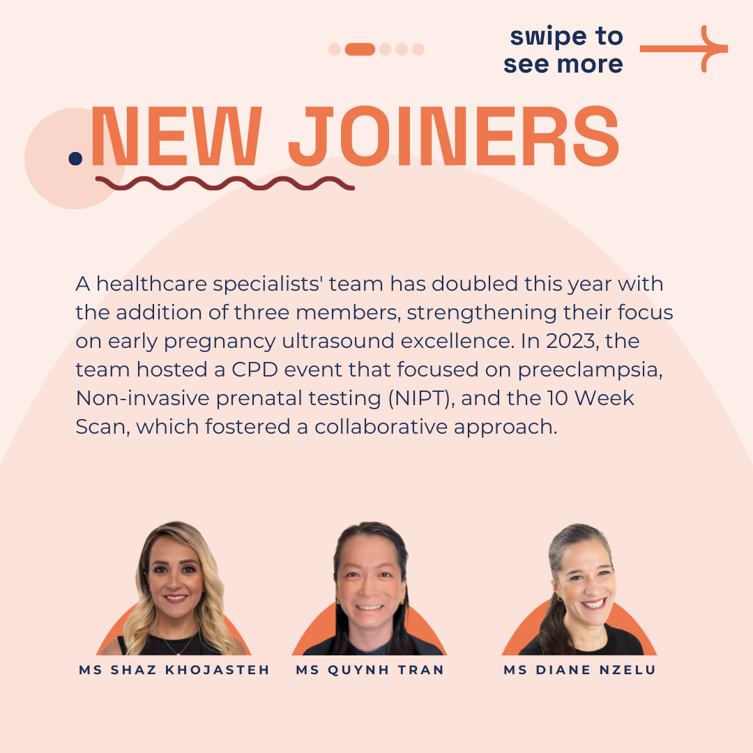
This year, our healthcare specialists’ team impressively doubled, boosting our capacity for expert care. We welcomed Ms. Shaz Khojasteh and, in December, Ms. Diane Nzelu. Their expertise has been invaluable, enhancing our mission of early pregnancy ultrasound excellence.
2023 marked our inaugural CPD event at Wallacespace. We delved into preeclampsia, Non-invasive prenatal testing (NIPT), and the 10 Week Scan. These sessions bolstered our collaborative approach, all the while maintaining a focus on early pregnancy ultrasound excellence.
Revolutionising Early Pregnancy Scans and NIPT
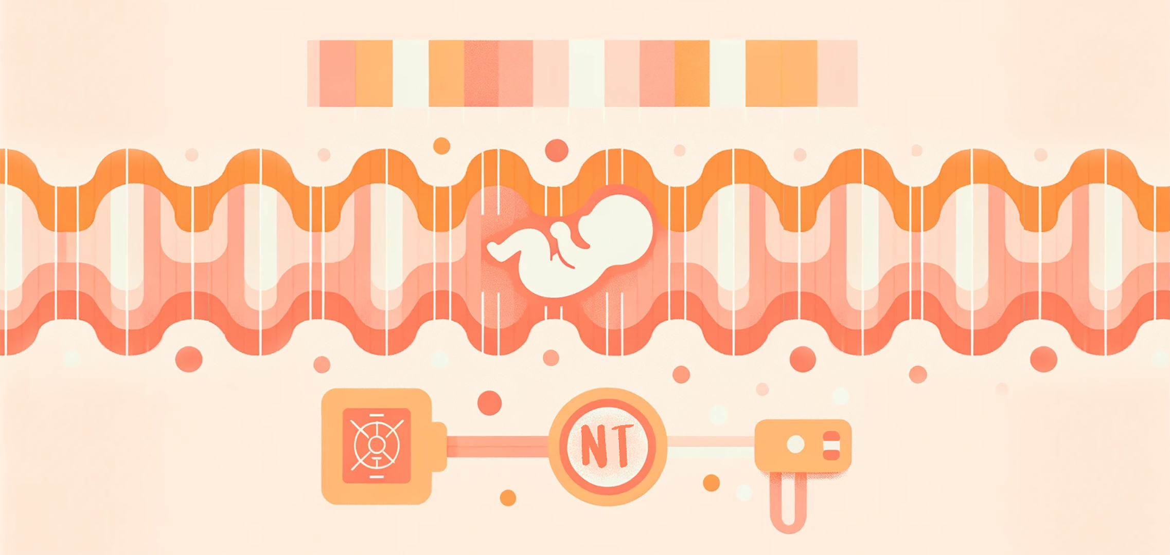
Significant changes marked our NIPT services this year. With Harmony’s closure, we partnered with Unity and PrenatalSafe. These alliances ensure that our patients continue to receive the utmost in screening technologies, reinforcing our commitment to early pregnancy ultrasound excellence. As we move forward, our commitment extends beyond providing current services; we are dedicated to researching and adopting the best NIPTs available. This commitment includes not only the screening’s accuracy and comprehensiveness but also the logistical aspects like delivery and result wait times, ensuring that our services are as convenient as they are reliable.
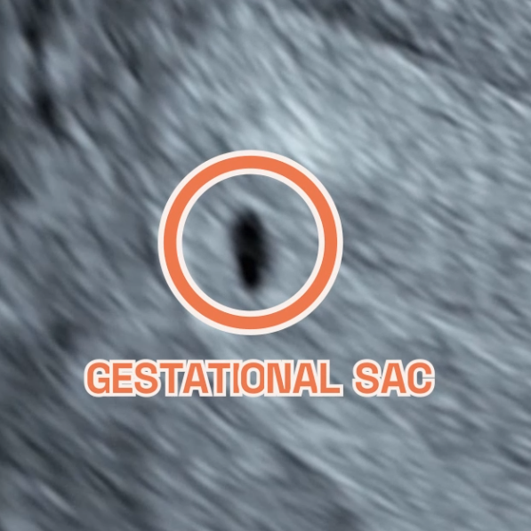
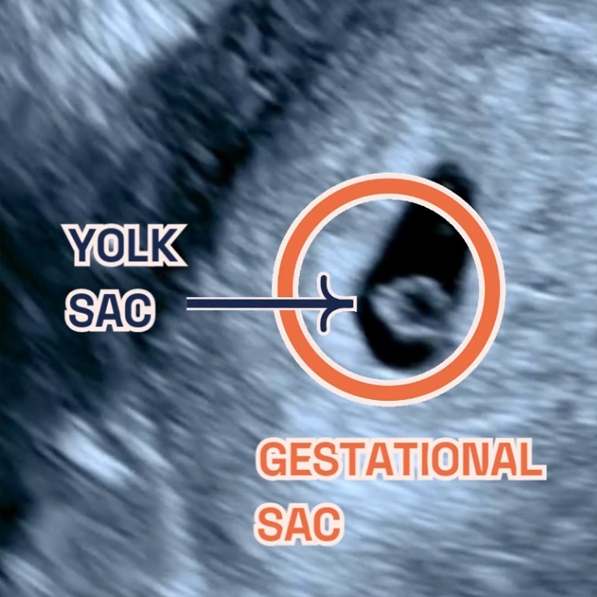
This year, we launched the ‘Smart Test‘, innovating early pregnancy diagnostics. This comprehensive product utilises a dual approach of early ultrasound, epitomising our commitment to early pregnancy ultrasound excellence. As we continue to embrace and refine this service, we are excited to announce that 2024 will bring even more detailed discussions and insights about the ‘Smart Test’.
We plan to delve deeper into how this test is transforming early pregnancy care, sharing the success stories and the science behind it. We’ll provide more educational content, including webinars, articles, and videos, to help expectant mothers and families understand the value and implications of the ‘Smart Test’. Our team of experts will also be available to answer questions, address concerns, and discuss how this innovative approach is part of the future of prenatal care.
Our focus has been on integrating NIPTs that offer a blend of high detection rates, low false positives, and a wide range of detectable conditions. We understand that the waiting period for results can be an anxious time for expectant parents. Thus, we are actively working to reduce this duration by streamlining processes and engaging with faster, yet equally reliable, lab services.
We aim to make NIPTs more accessible to a wider range of our patients, understanding that early and accurate screening can make a significant difference in prenatal care and planning. We are investigating more cost-effective solutions without compromising quality, aiming to include as many expectant parents as possible in the benefits of advanced prenatal screening.
Engaging Community Online
Remarkably, our dedication to educating and engaging with our community translated into significant online milestones. We reached 19,000 subscribers on YouTube, a testament to our engaging and informative content. Each video is a part of our commitment to demystify pregnancy and provide accessible, expert advice. From tips for a healthy pregnancy to detailed explanations of prenatal tests, our YouTube channel has become a valuable resource for expectant mothers worldwide.
Additionally, we received over 300 reviews on Trustpilot, reflecting the trust and satisfaction of our patients. These reviews are more than numbers; they are stories of the personal, positive impacts our services have had. Each review motivates us to continue improving and reaffirms our commitment to providing the highest standard of care.
Conclusion
In reflection, 2023 has been a year of solidifying foundations and embracing growth. Our team’s dedication has been unwavering, fuelled by a shared vision of excellence. Furthermore, our partnerships have strengthened, and our community has grown, all united by the goal of superior prenatal care.
As we look forward to 2024, we are filled with hope and anticipation. Our plans are ambitious, and aimed at further enhancing our services and outreach. Additionally, we remain dedicated to our educational initiatives, understanding their role in empowering expectant mothers.
In sum, 2023 has been a stepping stone, laying the groundwork for a future bright with possibility. We thank each member of our community for their part in this journey. Together, we look forward to continuing our commitment to early pregnancy ultrasound excellence in the coming year.