
Tag: hycosy

HyCoSy vs HSG: Which Fallopian Tube Test Is Right for You

Medical ultrasound equipment used for HyCoSy vs HSG fallopian tube testing comparison

HyCoSy vs HSG: Which Fallopian Tube Test Is Right for You

Published

Tags

Fertility investigations affect approximately 1 in 7 couples in the UK, and understanding which fallopian tube test is most suitable for your circumstances can be crucial for your journey towards conception. Two primary diagnostic procedures, HyCoSy and HSG, offer different approaches to assessing fallopian tube patency and uterine health, each with distinct advantages and considerations that may influence your choice.

Understanding Fallopian Tube Testing

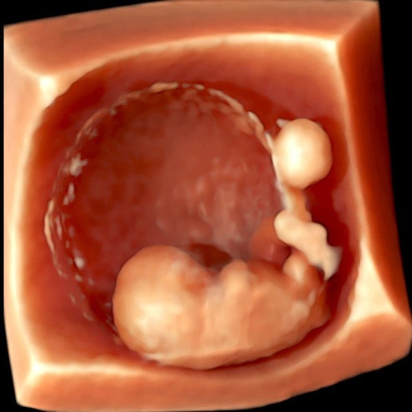
Fallopian tube testing forms an essential component of fertility assessment, helping clinicians identify potential blockages or abnormalities that may prevent conception. These diagnostic procedures evaluate whether eggs can travel freely from the ovaries through the fallopian tubes to reach the uterus, where fertilisation and implantation occur.

The two most commonly used non-surgical methods are HyCoSy (Hysterosalpingo-Contrast Sonography) and HSG (Hysterosalpingography). Both procedures involve introducing contrast medium into the reproductive system to visualise the internal structures, but they utilise different imaging technologies to achieve this assessment.

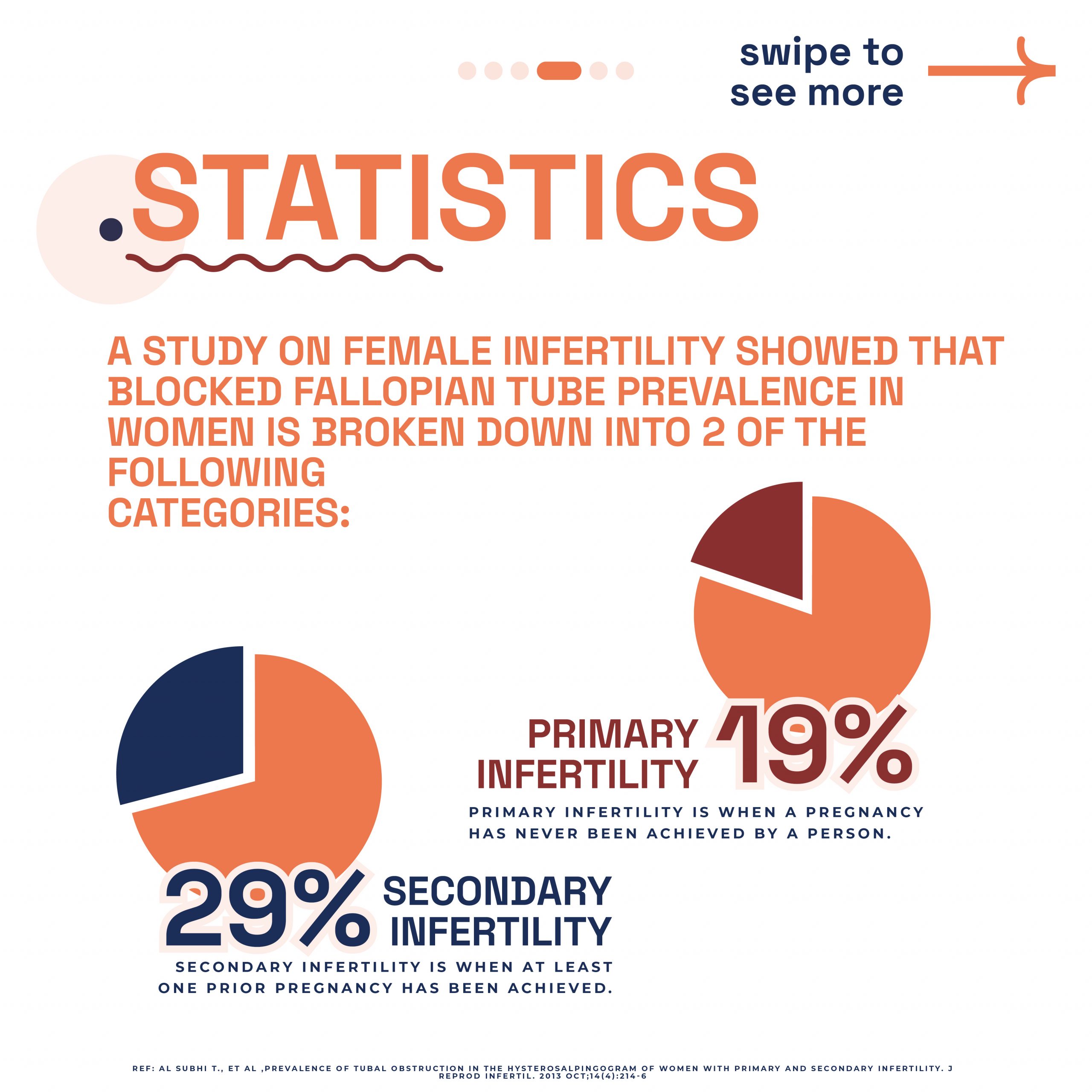
According to NHS guidelines, tubal factor infertility accounts for approximately 25-35% of female fertility issues, making accurate assessment of fallopian tube function vital for developing appropriate treatment plans.

What is HyCoSy

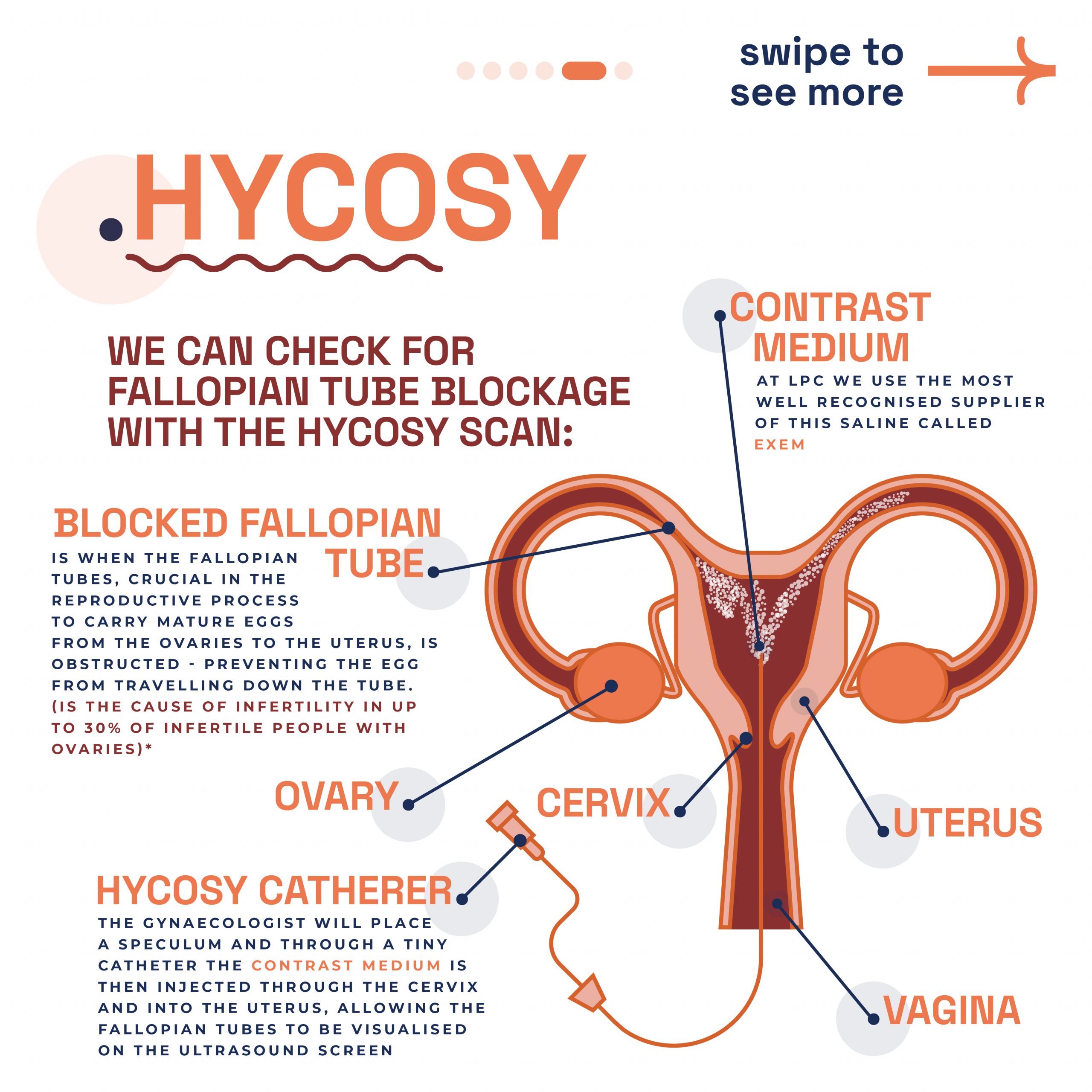
HyCoSy, or Hysterosalpingo-Contrast Sonography, represents a modern ultrasound-based approach to fallopian tube assessment. During this procedure, a specialised contrast medium is introduced through the cervix into the uterine cavity whilst real-time ultrasound monitoring captures detailed images of the reproductive organs.

The HyCoSy procedure utilises high-frequency sound waves to create detailed visualisations of both the uterine cavity and fallopian tubes. The contrast medium appears bright white on the ultrasound screen, allowing clinicians to observe its flow through the reproductive system and identify any areas of blockage or abnormal anatomy.

This technique offers the advantage of being radiation-free, making it particularly suitable for women who prefer to avoid X-ray exposure or who may require repeated assessments during their fertility journey.

What is HSG

HSG, or Hysterosalpingography, represents the traditional gold standard for fallopian tube assessment, utilising X-ray technology to visualise the reproductive organs. During this procedure, a radio-opaque contrast dye is introduced into the uterine cavity through the cervix, with X-ray images captured to show the dye’s progression through the fallopian tubes.

The HSG procedure provides excellent image quality and has been extensively studied and validated over decades of clinical use. The X-ray images offer exceptional detail of the uterine cavity shape and can clearly demonstrate whether the fallopian tubes are patent (open) or blocked.

HSG has traditionally been considered the first-line investigation for tubal patency assessment in many fertility clinics, with extensive research supporting its accuracy and reliability in diagnosing various reproductive conditions.

Key Differences Between HyCoSy and HSG

Understanding the fundamental differences between these two procedures can help you make an informed decision about which test may be most appropriate for your circumstances:

  • Imaging Technology: HyCoSy uses ultrasound waves whilst HSG employs X-ray radiation to create images of the reproductive organs.
  • Radiation Exposure: HyCoSy involves no radiation exposure, whereas HSG uses a controlled amount of ionising radiation.
  • Real-time Monitoring: HyCoSy provides real-time visualisation during the procedure, allowing immediate assessment of contrast flow.
  • Contrast Medium: HyCoSy uses specialised ultrasound contrast agents, whilst HSG employs radio-opaque dye visible on X-ray.
  • Procedure Duration: Both procedures typically take 15-30 minutes, though HyCoSy may be slightly quicker due to real-time imaging capabilities.
  • Equipment Requirements: HyCoSy requires specialised ultrasound equipment, whilst HSG needs X-ray facilities and fluoroscopy capabilities.

Research indicates that both procedures demonstrate comparable accuracy in detecting tubal blockages, with studies showing similar sensitivity and specificity rates for identifying fallopian tube abnormalities.

Advantages of HyCoSy

HyCoSy offers several compelling advantages that make it an attractive option for many women undergoing fertility assessment:

  • No Radiation Exposure: The absence of ionising radiation makes HyCoSy suitable for women who wish to avoid X-ray exposure or require multiple assessments.
  • Real-time Assessment: Immediate visualisation allows clinicians to observe contrast flow dynamics and make instant assessments during the procedure.
  • Comfortable Environment: The procedure can be performed in a standard ultrasound room rather than requiring specialised X-ray facilities.
  • Detailed Ovarian Assessment: Ultrasound technology allows simultaneous evaluation of ovarian morphology and other pelvic structures.
  • Reduced Contrast Requirements: Often requires smaller volumes of contrast medium compared to HSG procedures.
  • Immediate Discussion: Results can be discussed immediately after the procedure, reducing anxiety and waiting times.

Clinical studies have demonstrated that HyCoSy provides excellent visualisation of uterine cavity abnormalities and can effectively identify both proximal and distal tubal blockages with high accuracy rates.

Advantages of HSG

Despite being the more traditional approach, HSG continues to offer distinct advantages in certain clinical situations:

  • Established Gold Standard: Decades of clinical use and extensive research validation support HSG’s reliability and accuracy.
  • Superior Image Quality: X-ray technology often provides exceptional detail of uterine cavity shape and tubal anatomy.
  • Widely Available: HSG facilities are commonly available in most hospitals and fertility centres across the UK.
  • Detailed Documentation: X-ray images provide permanent, high-quality records for future reference and specialist consultations.
  • Cost-Effective: HSG may be slightly more cost-effective in some healthcare settings due to established protocols and equipment.
  • Therapeutic Benefits: Some studies suggest that HSG may have mild therapeutic effects, potentially improving conception rates in certain cases.

The RCOG recognises HSG as an effective method for assessing tubal patency, with guidelines supporting its use as part of comprehensive fertility investigations when clinically appropriate.

Potential Disadvantages and Considerations

Both procedures have specific limitations and considerations that may influence your decision:

HyCoSy Considerations:

  • Operator Dependency: Results may vary depending on the sonographer’s experience and expertise with the technique.
  • Image Quality Limitations: Ultrasound images may be affected by patient factors such as body habitus or bowel gas.
  • Specialised Equipment: Requires specific ultrasound contrast agents and experienced personnel trained in the technique.
  • Limited Availability: May not be available in all healthcare facilities compared to traditional HSG.

HSG Considerations:

  • Radiation Exposure: Involves controlled X-ray exposure, which some women prefer to avoid.
  • Contrast Reactions: Rare but possible allergic reactions to iodine-based contrast media.
  • Facility Requirements: Requires access to specialised X-ray equipment and trained radiological staff.
  • Static Images: Provides snapshot images rather than real-time dynamic assessment of contrast flow.

Procedure Experience and Comfort

Understanding what to expect during each procedure can help you prepare mentally and physically for the assessment:

HyCoSy Experience: The procedure takes place in a comfortable ultrasound room, similar to routine pregnancy scans. You’ll lie on an examination couch whilst a transvaginal ultrasound probe and small catheter are used to introduce contrast and monitor its flow. Most women describe the experience as similar to a smear test with mild cramping during contrast injection.

HSG Experience: The procedure occurs in an X-ray department, where you’ll lie on an examination table beneath X-ray equipment. A speculum is used to visualise the cervix, and contrast dye is introduced through a small catheter. The experience may involve slightly more intense cramping, particularly when the contrast fills the fallopian tubes.

Both procedures are typically performed without anaesthesia, though some clinics may offer pain relief options. Most women can return to normal activities immediately after either procedure, though mild cramping or light spotting may occur for 24-48 hours.

Accuracy and Clinical Outcomes

Research comparing HyCoSy and HSG demonstrates that both procedures offer comparable accuracy in detecting tubal abnormalities. Clinical studies indicate sensitivity rates of approximately 85-95% for both techniques in identifying blocked fallopian tubes, with specificity rates similarly high.

A systematic review published in fertility journals found no significant difference between HyCoSy and HSG in terms of diagnostic accuracy for tubal patency assessment. Both procedures effectively identify:

  • Tubal Blockages: Complete or partial obstruction of one or both fallopian tubes.
  • Uterine Abnormalities: Structural anomalies affecting the uterine cavity shape or size.
  • Hydrosalpinx: Fluid accumulation within blocked fallopian tubes.
  • Adhesions: Scar tissue affecting tubal function or mobility.

The choice between procedures often depends on individual circumstances, patient preferences, and clinical considerations rather than significant differences in diagnostic accuracy.

Timing and Preparation

Both HyCoSy and HSG require careful timing and preparation to ensure optimal results and patient safety:

Optimal Timing: Both procedures should be performed in the first half of your menstrual cycle, typically between days 7-12, to avoid potential early pregnancy and ensure the endometrium is appropriately thin for clear visualisation.

Pre-procedure Preparation:

  • Contraception: Reliable contraception should be used from the start of your cycle until the procedure to prevent pregnancy.
  • Infection Screening: Some clinics may require screening for sexually transmitted infections before the procedure.
  • Pain Management: Taking ibuprofen 1-2 hours before the procedure may help reduce discomfort.
  • Medical History: Inform your clinician about any allergies, particularly to contrast media or medications.

Your healthcare provider will provide specific preparation instructions tailored to your individual circumstances and the chosen procedure.

Cost Considerations and NHS Availability

The availability and cost of HyCoSy versus HSG can vary significantly depending on whether you choose NHS or private healthcare pathways:

NHS Provision: Both procedures may be available through NHS fertility services, though availability varies by region and local commissioning decisions. HSG tends to be more widely available across NHS trusts, whilst HyCoSy availability may be limited to specialist centres.

Private Healthcare: Private fertility clinics typically offer both options, with costs varying depending on location and clinic facilities. HSG may be slightly less expensive in some settings due to established protocols and wider availability of equipment.

When considering costs, factor in potential follow-up appointments, additional imaging requirements, and the overall fertility assessment pathway rather than just the individual procedure cost.

Making Your Decision

Choosing between HyCoSy and HSG involves considering multiple factors specific to your individual circumstances:

  • Radiation Sensitivity: If you have concerns about radiation exposure or require multiple assessments, HyCoSy may be preferable.
  • Previous Procedures: Your experience with ultrasound scans versus X-ray procedures may influence your comfort level.
  • Clinical Complexity: Your clinician may recommend one procedure over another based on your specific medical history.
  • Availability and Timing: Local availability of equipment and expertise may influence your options.
  • Personal Preferences: Your comfort with different types of medical procedures and imaging technologies.

Discussing these factors with your healthcare provider will help determine which procedure aligns best with your individual needs and circumstances. Both procedures provide valuable diagnostic information to guide your fertility treatment journey.

How the London Pregnancy Clinic Can Help

At the London Pregnancy Clinic, we offer comprehensive HyCoSy scanning services performed by experienced sonographers using state-of-the-art ultrasound equipment. Our team understands the emotional and physical aspects of fertility investigations and provides compassionate, professional care throughout your assessment.

Our HyCoSy procedures are conducted in comfortable, private facilities with immediate results discussion and detailed reporting for your healthcare providers. We work closely with fertility specialists and can provide comprehensive imaging assessments as part of your broader fertility investigation pathway.

For women requiring additional screening or genetic counselling as part of their fertility journey, we also offer carrier screening tests and genetic counselling services to provide comprehensive reproductive health assessments.

Final Thoughts

Both HyCoSy and HSG represent valuable diagnostic tools in fertility assessment, each offering distinct advantages depending on your individual circumstances. The choice between these procedures should be based on careful consideration of your medical history, personal preferences, and clinical recommendations from your healthcare team.

Remember that these investigations form just one component of comprehensive fertility assessment, and the results will help guide your treatment options and reproductive health decisions. Whether you choose HyCoSy or HSG, both procedures provide crucial information about fallopian tube function and uterine health.

If you’re considering fallopian tube assessment as part of your fertility journey, our experienced team at the London Pregnancy Clinic is here to provide expert care and support. Contact us today to discuss your options and book a consultation to determine the most appropriate diagnostic pathway for your individual needs.

Latest Stories

Continue reading

Hertility x LPC partnership

Minimalist flat design illustration of fertility and pregnancy ultrasound partnership between specialists London Pregnancy Clinic and Hertility.

Hertility x LPC partnership:

Bringing Fertility and pregnancy ultrasound support for women

  • Published

  • Last Modified

Tags

Discover the transformative partnership between Hertility and London Pregnancy Clinic, revolutionising the way you access fertility and pregnancy ultrasound services. From early pregnancy to specialised scans, our collaboration ensures you receive world-class care without the wait, at a price you can afford.

In an exciting development for expectant mothers and those on their fertility journey, Hertility has partnered with us, London Pregnancy Clinic (LPC), transforming access to reproductive healthcare. Understanding the emotional toll of waiting for your first pregnancy scan, we’ve come together to offer you world-class scanning facilities, ensuring early reassurance through ultrasound without the long wait. It promises to bridge the gap between fertility understanding and pregnancy care, offering an integrated approach to women’s health that is both convenient and comprehensive. Both fertility and pregnancy ultrasound are very important and we are going to unpack what we can offer.

Who is Hertility?

In 2020, Hertility was conceived with a distinct vision by Dr. Helen O’Neill, Dr. Natalie Getreu, and Deirdre O’Neill. It sprang from a realisation that women’s health complexities were often overlooked, with conditions remaining enigmatic within the healthcare system. Hertility emerged as a beacon, championing the cause of bringing reproductive science into the hands of women globally.

Tired of the gaps in women’s healthcare, Hertility was born out of necessity. It became clear that the data underpinning women’s health was not only insufficient but also largely based on male physiology, leading to a paradigm shift in approach. Dr. O’Neill, through her fifteen years in academia, recognised the urgency for tailored datasets that reflect the true nature of female health concerns. Hertility is not just a company; it’s a movement towards empowering women with the knowledge to make informed decisions about their health and life choices.

Why was Hertility Created?

The inception of Hertility was motivated by the profound desire to address the elephant in the womb – the inherent complexities of female reproductive health. With a staggering 60% of women grappling with hormone and gynae issues and the diagnosis of fertility disorders stretching up to eight years, Hertility’s founders saw the dire need for change. By empowering women with accessible, research-led health assessments, Hertility is breaking barriers and shining a light on the path to understanding and managing women’s health effectively.

Hertility’s resolve to be research-led has set a new standard in women’s healthcare. It stands firmly on the foundation of clinical trials and extensive data collection, ensuring every insight provided is robust and actionable. Hertility’s comprehensive at-home hormone and fertility tests are more than just diagnostics; they are a clarion call to acknowledge and act on women’s reproductive health needs. The company’s commitment to sharing their findings reflects their ethos of transparency and education, making Hertility a true vanguard in the realm of women’s health.

Why Hertility and LPC?

Hertility is at the forefront of women’s health, offering at-home hormone and fertility test kits designed to empower women with knowledge about their reproductive health. Their comprehensive approach evaluates various factors, including hormonal balance, ovarian reserve, and potential reproductive health conditions, to provide actionable insights.

Understanding the importance of seamless care, Hertility’s partnership with LPC is a natural progression. LPC’s reputation for excellence in fertility and pregnancy ultrasound complements Hertility’s mission, providing a continuum of care that supports women from fertility assessment through to pregnancy. From fertility screening to anomaly scan we provide a large variety of services for women from our London clinic.

  • Fertility and pregnancy ultrasound services at  London Pregnancy Clinic - Viability scan

    Viability scan 3D image

  • Image showing what would an ultrasound show at 6 weeks of pregnancy. You can see the fetal pole, gestational sac and yolk sac. It shows an Ultrasound image by London Pregnancy Clinic that shows the gestational sac and how small it is - for educational purpose.

    You can see the fetal pole, gestational sac and yolk sac at week 6.

Fertility and pregnancy ultrasound

  • Early Pregnancy Scans: Available from 6 weeks, these scans offer early reassurance, confirming the presence of a gestational sac and fetal heartbeat. The first scan we offer is called the viability scan (or the 6-week scan).
  • HyCoSy Scans: For those investigating fertility concerns, HyCoSy scans assess tubal patency, crucial for understanding potential fertility issues.
  • General, Anomaly, and Early Fetal Scans: A comprehensive range of scans ensures that every aspect of your pregnancy is monitored, from general health checks to detailed anomaly scans. We specialise in NIPT and 10 Week Scan, which is our signature early pregnancy scan.

All scans at LPC are conducted by consultant-level doctors or specialist sonographers in fetal medicine, gynaecology, and fertility. Overseen by Dr Fred Ushakov, a renowned specialist in fetal medicine and obstetrical ultrasound, our team guarantees the highest standard of care.

Why Wait? Early Scans Available from 6 Weeks

The first few weeks of pregnancy are a profound journey filled with anticipation and, sometimes, anxiety. Traditionally, expecting mothers wait until the 11 to 14-week mark for their first scan, a period filled with unanswered questions and needless worries. Recognising the importance of early reassurance, LPC opens its doors to Hertility customers for pregnancy scans starting as early as 6 weeks. Our goal? To provide you with the peace of mind that comes from knowing your pregnancy is progressing as expected, as soon as possible.

If you’re navigating the journey of fertility and pregnancy, let Hertility and LPC guide you to a path of informed, supported, and comprehensive care. You can check out our fertility and pregnancy ultrasound services!

Final thoughts

Hertility’s partnership with the London Pregnancy Clinic marks a defining moment in women’s health. With Hertility’s research-driven methodology and our fertility and pregnancy ultrasound services, we hope to provide a holistic approach for all women. Our combined effort provides an answer to the calls for early and accessible pregnancy scans, ensuring that expectant mothers receive the support and assurance they need, precisely when they need it. This collaboration goes beyond convenience; it is a powerful statement that women’s health deserves prioritisation and respect.

Latest Stories

Continue reading

What is a Fallopian Tube?

Illustration detailing the expert fertility assessment of fallopian tubes offered at London Pregnancy Clinic.

Fallopian Tubes: all you need to know

Discovering the importance of fallopian tubes for fertility.

  • Published

  • Last Modified

Tags

Explore the critical role of fallopian tubes, or uterine tubes, in pregnancy and fertility. This post delves into their anatomy, function, and common issues affecting fertility, providing essential insights for expectant mothers and those planning pregnancy.

Welcome to our comprehensive guide on the fallopian tubes, an essential component of the female reproductive system. We also explore common pathologies that affect these tubes, leading to infertility issues and complications like ectopic pregnancies. Our esteemed in-house gynae team, brings their extensive knowledge and expertise to this discussion, providing invaluable insights into the significance of maintaining fallopian tube health. Join us as we unfold the mysteries of the fallopian tubes and their vital function in the miracle of life.

What are Fallopian Tubes

In the female body, the pelvis houses two vital structures known as fallopian tubes. One on either side of the uterus. These tubes act as pathways, connecting the ovaries to the uterus. Each fallopian tube is made up of four distinct parts: the infundibulum, fimbriae, ampulla, and isthmus. The fimbriae are delicate, finger-like extensions that gently guide the egg from the ovary into the tube. 

The Role of Fallopian tubes in Pregnancy

Moreover, fallopian tubes are critical in the earliest stages of pregnancy, integral to the reproductive process, and serve a trio of critical functions. They are the conduits for egg transportation from the ovaries to the uterus and the venue where fertilisation typically occurs. Post-fertilisation, these tubes facilitate the journey of the fertilised egg to the uterus, setting the stage for implantation and the progression of pregnancy. The health and integrity of the fallopian tubes are paramount for successful conception as they are closely linked to fertility. Problems with these tubes are a common reason why some women may have difficulty getting pregnant.

Uterine Tube Pathologies & Infertility

According to a report, nearly one-third of infertility cases are attributed to issues with the fallopian tubes, including various pathologies like inflammation and obstructions. Damage to the tube’s cilia can hinder the movement of sperm or egg, contributing to infertility. Additionally, sexually transmitted infections are a known cause of these tubal issues. Let’s dive into the specific pathologies that can manifest.

Salpingitis

Salpingitis, an inflammation in the fallopian tubes, often forms part of wider pelvic inflammatory diseases (PIDs). Its hallmark is a thickened tube in the isthmus area, known as salpingitis isthmica nodosa. This condition can resemble endometriosis, potentially leading to fertility blockages or raising ectopic pregnancy risks.

During diagnosis, doctors first look for tenderness and swelling. They may conduct blood and urine tests to spot infection indicators. Swab tests from the vagina and cervix help identify specific bacterial infections. Ultrasound scans of the fallopian tubes and reproductive tract are common. A crucial test, the hysterosalpingogram, uses a special X-ray and dye through the cervix to detect tubal blockages.

Untreated, salpingitis can cause long-term fertility issues and increase ectopic pregnancy chances. Symptoms vary, including lower abdominal pain, nausea, and fever; sometimes, no symptoms appear initially. Antibiotics typically treat this condition, but severe cases might require surgery or IVF. Seeking medical advice early is vital to prevent serious outcomes. Early detection and treatment significantly aid in managing salpingitis effectively.

Fallopian Tube Blockage & Narrowing

Addressing fallopian tube obstructions can significantly boost pregnancy chances. These blockages may develop in various parts of the tube – proximal, distal, or mid-segmental. Testing the tubes’ full functionality presents challenges, yet assessing their patency (openness) is possible with hysterosalpingography, laparoscopy with dye, or hysterosalpingo contrast sonography (HyCoSy). During surgical evaluations, specialists examine the tubes and introduce a dye, such as methylene blue, into the uterus to test its passage through the tubes when the cervix is closed.

Research published in PubMed indicates a notable prevalence of tubal obstruction: 19.1% in primary infertility cases and 28.7% in secondary infertility scenarios. The study highlights cornual blockage’s predominance in primary infertility, whereas complete hydrosalpinx and peritubal adhesions appear less frequently. Due to the frequent link between tubal disease and Chlamydia infection, Chlamydia antibody screening is now a cost-effective approach for identifying possible tubal pathologies.

At London Pregnancy Clinic, we conduct the HyCoSy procedure. This test evaluates fallopian tube functionality by injecting a special dye and monitoring its flow. It also assesses the womb for abnormalities affecting pregnancy implantation. Additionally, we use dye and ultrasound scans to check for fallopian tube blockages or abnormalities. Discover more through the slider below, offering further insights into our advanced diagnostic techniques.

  • Graphic explaining the expert fertility assessment of fallopian tubes offered at London Pregnancy Clinic.

    Fallopian Tubes

  • Educational diagram of female reproductive anatomy focusing on the fallopian tubes provided by London Pregnancy Clinic.

    Fallopian tube diagram

  • Illustration detailing the phases of the menstrual cycle as part of fertility education by London Pregnancy Clinic.

    Menstrual Cycle

  • Infographic showing statistics on blocked fallopian tube prevalence in women, as part of London Pregnancy Clinic's fertility resources.

    Stats on blocked fallopian tube

  • Explanation of the HYCOSY scan procedure for checking fallopian tube blockage, offered by London Pregnancy Clinic.

    HyCoSy

  • Image of Mr. Prashant Purohit with text highlighting London Pregnancy Clinic's expertise in fertility and gynaecology ultrasound.

    Mr. Prashant Purohit specialist gynaecologist

Ectopic Pregnancy

Statistics and Locations of Ectopic Pregnancies: Ectopic pregnancies, which occur in about 1%-2% of all pregnancies, predominantly implant in the fallopian tubes, with over 98% of cases occurring there. Within the fallopian tubes, the distribution of ectopic pregnancies varies: approximately 75% happen in the ampullary segment, 13% in the isthmic segment, and around 12% in the fimbrial segment. These statistics highlight the critical nature of monitoring tubal health.

Fertilisation and its Relation to Ectopic Pregnancies: It’s important to note that fertilisation typically occurs in the ampulla, the same section of the fallopian tube where the majority of ectopic pregnancies are found. This correlation emphasises the need for awareness and early detection in cases of suspected ectopic pregnancy, as the ampulla is a common site for both normal fertilisation and ectopic implantation.

Understanding the statistics and locations of ectopic pregnancies in the fallopian tubes is crucial. It not only sheds light on the commonality and risks associated with these pregnancies but also underscores the importance of early ultrasound pregnancy scans for detection and intervention. The earlier you scan the quicker we can detect an ectopic pregnancy meaning that we can begin this from week 6 of pregnancy.

Pregnant with Just One Fallopian Tube?

Typically, women possess two fallopian tubes for transporting the mature egg from the ovary to the uterus, where it may encounter sperm. However, a single healthy tube can efficiently undertake this task. Remarkably, up to 85% of women aged 22-28 with only one tube successfully conceive within two years of trying, even following an ectopic pregnancy.

If you have a blocked tube due to scarring, trauma, or a previous ectopic pregnancy, your pregnancy prospects remain normal, considering other health factors. Loss of a fallopian tube might result from pelvic surgery due to infection, tumours, or a past ectopic pregnancy. In some instances, women are naturally born with just one tube. Still, pregnancy is achievable with one tube, provided you have a functioning ovary, you’re ovulating and the fallopian tube is in good health.

If you’re struggling to conceive or have a history of fallopian tube issues, it’s wise to consult your gynaecologist or a reproductive endocrinologist. They can offer a thorough evaluation to guide your next steps. Remember, one healthy fallopian tube can effectively support your journey to motherhood.

Conclusion

Mr. Prashant Purohit, our dedicated in-house gynaecologist at London Pregnancy Clinic, emphasises the need for awareness and timely medical consultation to address these concerns. His expertise in managing these conditions reflects our clinic’s commitment to providing comprehensive care and support to women facing fertility challenges. Understanding the complexities of the fallopian tubes is not just about medical knowledge; it’s about empowering women with the information they need to make informed decisions about their reproductive health. We encourage you, readers to stay vigilant about their gynaecological well-being and seek expert guidance when needed. Remember, early detection and treatment are key to overcoming obstacles on the path to motherhood.

Latest Stories

Continue reading