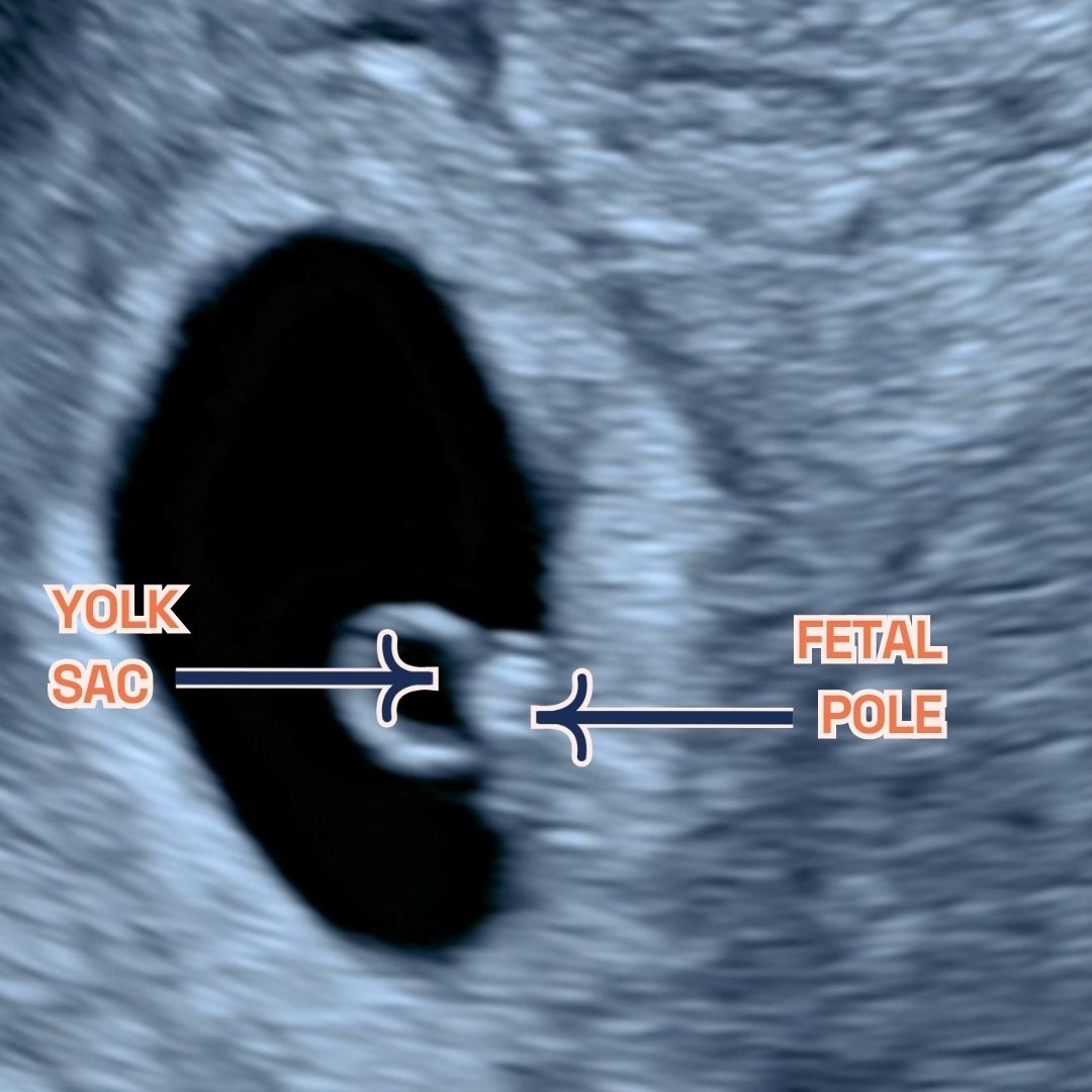
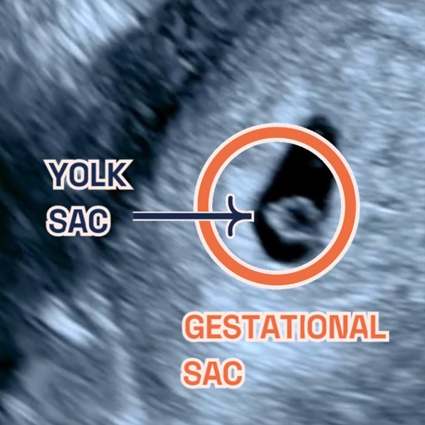
Tag: private 6 week scan

Understanding Pregnancy Tests

A modern, minimalist illustration depicting a hand holding a positive pregnancy test, surrounded by abstract shapes and elements in light orange and baby pink, symbolising joy and new beginnings.

Understanding Pregnancy Tests:

Your First Step Towards Motherhood and beyond.

  • Published

  • Last Modified

Tags

Discover the essentials of a pregnancy test, from how they work to the different types available and what steps to take after getting your results. This guide offers peace of mind as you embark on your journey to motherhood.

Pregnancy tests are the first beacon of information in your journey towards motherhood. They detect the presence of human chorionic gonadotropin (hCG), a hormone your body produces during pregnancy. Highly accurate, these tests offer reassurance and a preliminary confirmation of pregnancy, paving the way for the exciting journey ahead.

How does a Pregnancy Test Work?

Pregnancy tests search for hCG in your urine, which your body starts producing shortly after a fertilised egg attaches to your uterus. This hormone’s levels double every two to three days in early pregnancy, making urine and blood tests highly effective ways to confirm pregnancy. While home tests offer privacy and quick results, blood tests at your doctor’s office can confirm pregnancy earlier and more accurately.

Over-the-counter (OTC) tests detect the HCG hormone in urine, with various methods depending on the chosen kit. You might dip a stick into collected urine, use an eyedropper for transferring urine to a container, or place the stick directly in your urine stream mid-flow.

Home pregnancy tests boast a near-99% accuracy rate when used as directed by the Office on Women’s Health. These tests offer privacy, simplicity, and quick results, usually within minutes. Results could appear as a color change, a line, a symbol (like a plus or minus), or explicitly as “pregnant” or “not pregnant.” A negative outcome might not be definitive, especially if instructions weren’t followed, the test was taken too soon, or certain medications interfered. Consult a pharmacist about potential medication effects on test results.

If symptoms persist or your period doesn’t commence, try another test in a week. Seek GP advice for continuous negative results without menstruation. Some pharmacies and GPs provide tests, sometimes free or for a nominal fee, ensuring even quicker answers. Remember, if going to your GP, bring a urine sample in a clean container. Results can take days, though some tests are instant.

Wondering when to take a pregnancy test?

Some highly sensitive tests can detect pregnancy before your period is due. Yet, waiting until the first day of a missed period boosts accuracy. For the clearest result, test a week after missing your period.

Tests measure HCG, a hormone present only during pregnancy. This hormone appears after a fertilized egg attaches to the uterus, a process known as implantation, occurring about 10 days post-conception. HCG traces can appear 6 days post-implantation, but levels usually need 7–10 days to become detectable. If your cycles are irregular, testing 21 days after unprotected intercourse is wise. Testing too early may yield inaccurate results.

Types of Pregnancy Tests

Opt for Home Pregnancy Tests: Seeking a convenient option? Home pregnancy tests offer privacy. Conduct these tests as soon as you miss your period. They promise up to 99% accuracy when used as directed.

Consider Blood Tests: Need an early confirmation? Blood tests, performed in healthcare settings, can provide it. Although less commonly used, they’re highly accurate for early detection. At London Pregnancy Clinic we can perform these tests and one of our phlebotomists will assist you.

Signs you may be pregnant

Spotting early pregnancy signs is crucial. If you notice any unusual changes, consider a pregnancy test. Here’s a restructured and concise guide to understanding these signs.

Missed Period: A hallmark sign, missing your period suggests it’s time for a test. While a standard cycle lasts about 28 days, they can range from 24–38 days. If over a month has passed without your period, testing is wise. However, stress, medication, and changes in birth control can also affect your cycle. Early pregnancy might show light spotting as the egg implants, which differs from a regular period. Consult a doctor for any bleeding with a positive test.

Cramping: Similar to menstrual cramps, early pregnancy might bring discomfort. If your period doesn’t follow, testing is advisable.

Breast Changes: Pregnancy increases estrogen and progesterone levels, leading to tender, fuller breasts and darker veins. These symptoms can also precede a period, so they’re not definitive of pregnancy.

Feeling Different: Early signs include nausea, food aversions, fatigue, and frequent urination. Pay attention to your body; any distinct changes might indicate it’s time for a test.

Failed Birth Control: No method is 100% foolproof. Pills might fail, especially if not used perfectly, and condoms can break. Despite the effectiveness of long-term options like IUDs, pregnancy is still possible. If experiencing other pregnancy signs, take a test, regardless of your birth control use.

Remember, understanding your body’s signals is key. If you’re encountering any of these symptoms, a pregnancy test can provide early insight, allowing for prompt healthcare consultation. At London Pregnancy Clinic, we’re here to support you with tailored advice and comprehensive care for your pregnancy journey.

Embarking on Your Journey

A positive test marks the start of a remarkable journey. Your next move? Book an appointment with your healthcare provider. This crucial visit can verify your pregnancy with additional tests. It also kick-starts vital prenatal care, safeguarding your well-being and your baby’s health. Embrace this journey, prioritising health and care from the outset. Your provider will guide you through each step, ensuring you and your baby thrive.

At the LPC, we understand the emotional and physical significance of this phase in your life. Recognising the unique needs and concerns of expectant mothers, LPC offers the opportunity to book your first appointment directly with a midwife. This personal touch ensures that you receive tailored advice and support right from the outset.

Furthermore, LPC introduces an option that diverges from the standard NHS protocol. Recognising the desire for early reassurance, we offer a viability scan as early as 6-9 weeks into the pregnancy. This service caters to those eager for an earlier confirmation of their pregnancy’s progression, rather than waiting until the NHS’s 12-week scan. The decision to opt for this early scan underscores the importance of peace of mind and the value of early connection with the developing life within.

The viability scan presents a window into the womb, offering expectant parents their first glimpse of their unborn child and the heartbeat that symbolises life. This experience often strengthens the bond between parent and child, grounding the reality of the pregnancy and fostering an early emotional connection.

Ultrasound Screening Vital for Early Detection

Ultrasound screenings are not just routine checks; they’re crucial for early prenatal care. These scans help identify potential structural anomalies in the baby’s development, which are pivotal for addressing congenital anomalies. At London Pregnancy Clinic, we specialise in providing comprehensive ultrasound screenings, tailored to each stage of pregnancy, ensuring early detection and peace of mind from 10 weeks onward.

Many inquire about the possibility of conducting a scan at 4 weeks. However, at such an early stage, the ultrasound reveals limited details, often not enough to confirm a pregnancy. By 5 weeks, while we may observe the gestational and yolk sacs, these findings alone do not confirm pregnancy viability. It’s this understanding that shapes our recommendation for the optimal timing of your first scan to be held off until week 6/7.

Our 10 Week Scan stands out, offering a unique glimpse into the womb to detect 10 critical fetal anomalies. This pioneering scan, exclusive to our clinic, aligns perfectly with Non-Invasive Prenatal Testing (NIPT), elevating the standard of prenatal care available to you.

The cornerstone of our scanning services, the Early Fetal Scan, provides an in-depth examination aimed at identifying over 100 serious anomalies well before the standard NHS 19-20 week scan. This early insight grants expectant parents the reassurance and crucial information needed during these formative weeks.

Our approach to prenatal screenings is revolutionary, offering early answers and invaluable peace of mind. Through our advanced screening capabilities, we illuminate potential concerns, facilitating timely interventions. These scans are more than mere procedures; they are essential milestones providing reassurance that your baby’s development is on course, empowering you to manage your pregnancy with informed confidence and proactive care.

Final Thoughts

At the London Pregnancy Clinic, we’re here to support you from the very first step. If you’ve taken a pregnancy test and are ready for the next stage, or if you have questions about pregnancy tests, our team is here to assist you. You can use our EDD calculator to figure out when your baby might arrive.

Latest Stories

Continue reading

Baby Structures During Pregnancy

Graphic of a stork carrying a baby over the city of London hospital. Blog about baby structures.

Fetal Structures & Ultrasound:

Prenatal Ultrasound’s role in screening baby structures.

  • Published

  • Last Modified

Tags

  • Visual guide from London Pregnancy Clinic detailing ultrasound scans for baby structural development.

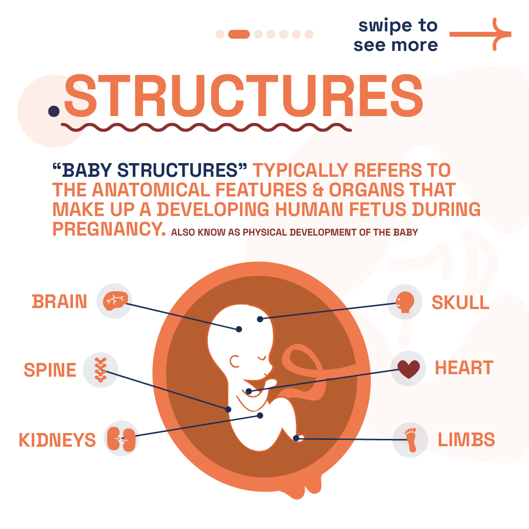
  • Detailed diagram from London Pregnancy Clinic showing key baby structures during pregnancy development.

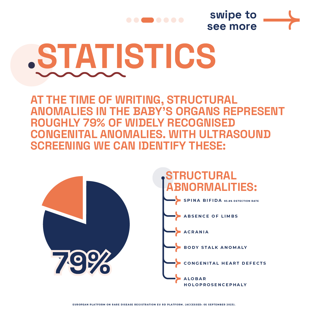
  • London Pregnancy Clinic statistics infographic on structural anomalies in fetal development with a 79% pie chart.

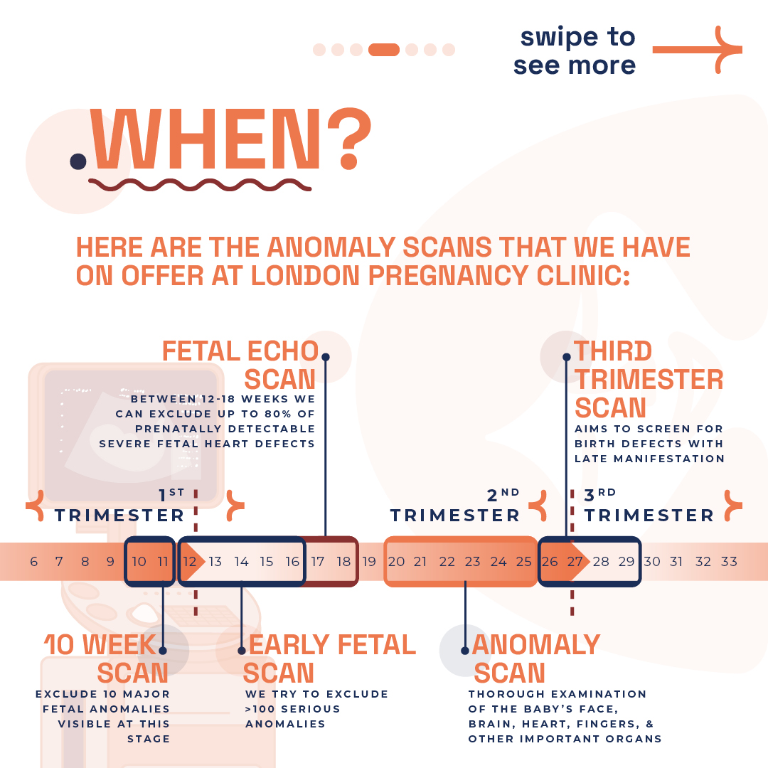
  • London Pregnancy Clinic timeline for fetal anomaly scans during pregnancy trimesters.

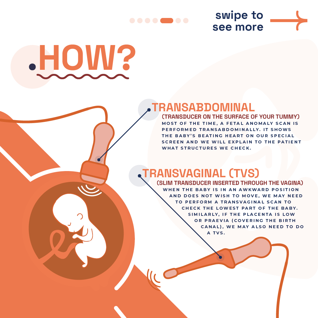
  • Illustration explaining transabdominal and transvaginal ultrasound scans at London Pregnancy Clinic.

  • Dr. Fred Ushakov at London Pregnancy Clinic highlighting expertise in fetal anomaly screenings.

  • FAQ section on London Pregnancy Clinic website discussing ultrasound scans for genetic anomalies detection.

Pregnancy is a journey of discovery, not just of the self but also of the little life growing inside you. Among the many wonders this period brings is the ability to glimpse into your baby’s developing world through ultrasound screenings. Here we delve into the critical aspects of baby structures, their development, and the pivotal role of ultrasound in ensuring the well-being of your unborn child.

As expectant mothers, the health and development of our unborn child is paramount. Advances in medical technology have gifted us with the ultrasound, a window into the womb, allowing us to monitor the growth and health of our babies like never before. Understanding the structures of a developing baby provides invaluable insights into their well-being and development.

What are fetal structures?

“Baby structures”, or fetal structures, usually denote the anatomical features and organs constituting a human fetus throughout pregnancy, also referred to as the physical development of the baby. It is important to note that this does not include genetic anomalies like Down Syndrome. Fetal development is a complex process where vital structures like the brain, heart, spine, and limbs evolve significantly:

Brain: Development kicks off around week 5 with the neural tube, leading to the brain and spinal cord’s formation. By the second trimester’s close, the brain’s structure mirrors that of an adult, showcasing an almost fully developed brain stem.

Heart: One of the earliest and most significant markers we observe during our dating scan is the heartbeat at 6 weeks, visible through ultrasound, providing crucial insight into the baby’s health and development at this early stage. This marks the beginning of the heart’s rhythmic life-sustaining beats.

Spine: The spine’s journey begins around weeks 6-7, laying the groundwork for the nervous system’s housing and body support. This period also sees the onset of bone development, integral to the body’s framework.

Limbs: Concurrently, weeks 6-7 see the emergence of arm and leg buds, with hands, feet, and digits evolving throughout the pregnancy. These developments are pivotal for the baby’s eventual movement capabilities.

The structures extend to encompass vital organs like the liver and kidneys, which are essential for processing nutrients and waste, the skull that protects the developing brain, and the umbilical cord, a lifeline connecting the baby to the placenta. Additionally, the abdominal organs, critical for digestion and other bodily functions, are part of this intricate network of development, underlining the complexity and wonder of fetal growth during pregnancy. Monitoring these structures’ growth is vital, employing fetal heart rate monitoring, ultrasounds, and physical exams to ensure the baby’s health and well-being.

Monitoring Key Baby Structures 

Expectant mothers cherish every moment of their pregnancy journey, closely observing their baby’s development. The fetus’s organs and anatomical features, notably the brain, heart, spine, and limbs, undergo remarkable transformations. These critical structures not only signify health but also ensure the baby’s overall development and well-being.

From the first to the third trimester, various scans are recommended to monitor your baby’s development. These range from the 10-week scan, aiming to exclude major fetal anomalies, to the fetal echo scan and the anomaly scan, each designed to screen for specific conditions at different stages of pregnancy .

The majority of fetal anomaly scans are conducted transabdominally (watch our video on ultrasound probes), offering a glimpse of the baby’s heartbeat and structures. In certain cases, a transvaginal scan might be necessary for a more detailed view, especially when the baby is positioned awkwardly or when examining the placenta.

As the fetus grows, so does our anticipation and concern for its health. Regular monitoring of these structures becomes paramount. It reassures us that development proceeds without any hindrances. Specifically, we focus on the baby’s heart and brain, pivotal for life and cognitive functions. The spine’s integrity is equally crucial, supporting the body and housing the nervous system. Moreover, observing the limbs ensures proper growth and the potential for movement. In essence, these structures form the foundation of our baby’s future. Ensuring their optimal development through vigilant monitoring not only secures their health but also paves the way for a thriving life post-birth.

Most Common Structural Anomalies?

According to the European platform on Rare disease registration EU RD Platform, structural anomalies in the baby’s organs represent roughly 79% of widely recognised congenital anomalies. The most common structural anomaly encountered in newborns is congenital heart defects (CHD). These defects, which disrupt the heart’s structure and function post-birth, affect 1 in 100 UK babies. Ventricular septal defect (VSD), or ‘hole in the heart,’ stands out as the predominant issue.

Newborns also face other frequent congenital issues, including:

  • Cleft Lip and Palate: A gap in the mouth’s upper lip and roof.
  • Spina Bifida: The spinal column’s incomplete closure.
  • Neural Tube Defects (NTD): Disorders like anencephaly affecting brain and spinal development. Spina bifida falls under NTDs.
  • Gastroschisis: Intestines extending outside the abdomen from birth.
    Exomphalos or Omphalocele: Abdominal organs bulge through the navel.
  • Clubfoot or Talipes: A foot deformity where it angles inwards and downwards.
  • Limb Reduction Defects: Absence or incomplete limb formation.
  • Polydactyly: Having extra fingers or toes.
  • Intestinal and Duodenal Atresia: Intestine blockages or missing sections.
  • Kidney Anomalies: Various kidney development, structure, or function malformations.

In essence, these conditions highlight the spectrum of potential challenges infants may face from birth, underscoring the importance of early detection and intervention. Awareness and understanding of these common anomalies can lead to better outcomes for affected children, as early intervention often opens the door to corrective procedures and supportive therapies, enhancing quality of life. The emphasis on these conditions reflects a commitment to providing the necessary care and support for affected families, guiding them through diagnosis, treatment, and beyond, with a focus on fostering resilience and hope.

Ultrasound Screening Vital for Early Detection

Ultrasound pregnancy scans transcend mere glimpses into the womb; they are indispensable tools in prenatal care. Structural anomalies within the baby’s organs account for a significant share of congenital anomalies. Fortunately, ultrasound offers a solution, detecting early signs of such conditions. At the London Pregnancy Clinic, our expertise in ultrasound screenings shines at every stage. We tailor our early pregnancy scans to the gestational age, offering unparalleled early detection from 10 weeks onwards.

10 Week Scan: Our clinic excels with a pioneering 10/11-week scan, designed to uncover 10 critical fetal anomalies early on. This scan, unique to our services, complements NIPT seamlessly, setting a new standard in prenatal care.

Early Fetal Scan: The hallmark of our service, the Early Fetal Scan, conducts an exhaustive review of the baby’s development. It’s crafted to spot over 100 serious anomalies well ahead of the standard NHS 19-20 week screenings. Offering this advanced insight provides early reassurance and essential information to expectant parents.

In short, our approach revolutionises prenatal screenings, giving you early answers and peace of mind. It brings to light potential issues, enabling timely interventions. These screenings are not just procedures but gateways to peace of mind for expectant mothers. They assure us that our baby’s development is on the right track, allowing us to address any concerns proactively.

  • Baby with missing left hand at 12 week early pregnancy scan.

    Baby with missing left hand at 12 week, an absence or incomplete limb formation.

  • Normal Early pregnancy scans at 12wk 3D.

    Normal baby at 12 weeks of pregnancy during an early pregnancy scans 3D.

Advanced Anomaly Detection in Late Pregnancy

Early detection of fetal anomalies enables timely interventions, crucial for the well-being of your baby. However, some anomalies manifest later, necessitating advanced screenings in the later stages of pregnancy. Our late-stage anomaly scans are designed to offer peace of mind and crucial insights during the final stages of pregnancy. 

Anomaly Scan: Our 22-23 week Anomaly Scan goes beyond the NHS’s 19–20-week standard, offering a deeper insight into your baby’s development. This comprehensive check includes detailed assessments of the brain, heart, face, fingers, and vital organs. We also provide additional services like uterine artery Doppler and cervical length measurements on request, ensuring an all-encompassing review.

3rd Trimester Anomaly Scan: To address the absence of routine NHS scans post-18-20 weeks, our clinic offers the 3rd Trimester Anomaly Scan between 26-29 weeks. This scan is pivotal for detecting late-manifesting anomalies and monitoring fetal growth and well-being. Given the technical difficulties of scanning after 30 weeks, this period is optimal for identifying significant issues, especially affecting the brain, heart, and kidneys. It’s also an ideal time for clinical 3D/4D imaging, enriching your connection with your unborn child.

Leveraging state-of-the-art technology and thorough assessments, we are committed to delivering unparalleled prenatal care. Trust our expertise to navigate you through these essential phases of pregnancy, providing clarity, early intervention opportunities, and fostering a profound bond with your baby.

Why do we focus on Early detection?

At our clinic, early detection of fetal anomalies stands as a priority, underlining our commitment to comprehensive prenatal care. Early diagnosis paves the way for detailed insights into potential health issues, fostering a deeper understanding and effective management from the outset.

This early intervention allows healthcare experts to convey critical information regarding the anomaly, its possible effects, and the spectrum of care and intervention options available. Such a proactive stance equips parents with the knowledge to make well-informed decisions, streamlines medical planning, and guarantees the availability of essential support and resources.

Given the time-sensitive nature of advanced genetic screenings, including microarrays and exome sequencing, alongside expanded NIPT choices, securing an early diagnosis is crucial. These intricate tests, demanding several weeks for results, underscore the necessity of timely identification. It ensures ample time for exhaustive testing, facilitating a holistic and well-considered approach to prenatal care and support throughout the pregnancy journey.

Embracing this early detection strategy, we aim to guide expectant parents with clarity, empowering them with options and support every step of the way. Our focus on early diagnosis reflects our dedication to delivering exceptional care and support, ensuring a well-informed, reassuring pregnancy experience.

Baby Structures FAQs


  • Can ultrasound detect genetic anomalies?

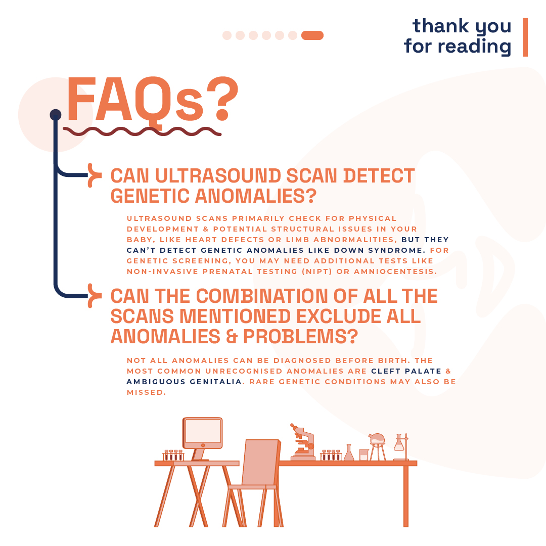
    While ultrasound scans are adept at identifying physical development and potential structural issues, they cannot detect genetic anomalies. For genetic screening, additional tests like Non-Invasive Prenatal Testing (NIPT) or amniocentesis are recommended.

  • Do the scans guarantee the detection of all anomalies?

    Regrettably, predicting every anomaly before birth is beyond reach. Commonly missed anomalies include isolated cleft palate (a split in the mouth’s roof with intact lips) and ambiguous genitalia. Additionally, certain rare genetic conditions elude detection through ultrasound scans.

  • Structural Anomalies Linked to Genetic Syndromes?

    Noticing structural anomalies or physical differences in a baby during pregnancy often hints at a genetic or chromosomal connection. Simply put, these anomalies might signal an underlying chromosomal or genetic syndrome, commonly referred to as “rare diseases.

    Understanding the tie between genetic makeup and structural anomalies is key. It unravels the complex relationship between a baby’s DNA and their physical traits. An anomaly might stand alone or indicate a broader genetic condition.

    If a baby is diagnosed with a chromosomal or genetic condition, these anomalies manifest the impact of irregular gene functions.

    Crucially, when structural defects accompany chromosomal or genetic conditions, the prognosis tends to be more serious than isolated anomalies. Such conditions frequently correlate with intellectual disabilities or significant health issues, underscoring the importance of early detection and intervention.

  • Why do NIPT with Scan and visa versa?

    Ultrasound plays a crucial role in fetal screening, essential for confirming viability and gestational age alongside NIPT submissions. While NIPT effectively screens for three chromosomal anomalies and other genetic conditions (specific to the panorama test), it doesn’t assess structural anomalies. Conditions like heart or brain defects, often more common and severe than Down’s syndrome, require ultrasound for detection.

    At the London Pregnancy Clinic, our forte is the early identification of fetal anomalies, particularly in the fetal heart and brain. Remarkably, we’re capable of detecting significant anomalies from as early as 10 weeks. Discover more about the scans we pair with NIPT by visiting our scans page; our 10-week scan frequently stands out as the optimum choice.

    Opting for a Panorama Test at 9 weeks? Rest assured, we’ll conduct a detailed structural scan of your baby, albeit with some limitations due to the embryo’s small size and early development stage.


Final Thoughts

Understanding the structures of your developing baby is a crucial part of the pregnancy journey. Ultrasound screenings offer an invaluable tool in this understanding, providing early detection of potential issues and contributing to the effective monitoring of your baby’s health. We invite you to experience the comprehensive care and advanced screening options available at the London Pregnancy Clinic, where we prioritise your peace of mind and your baby’s well-being.


Latest Stories

Continue reading

Week 9 of Pregnancy

Early pregnancy scan London Pregnancy Clinic illustration of week 9. Week 9 of pregnancy fetus.

Week 9 of your Pregnancy

Discovering your baby’s progress since week 8

  • Published

  • Last Modified

Tags

Week 9 marks a pivotal moment in your pregnancy journey, showcasing significant developments in your baby’s growth. London Pregnancy Clinic invites you to witness these fascinating changes through our private early pregnancy scans. This period highlights the transformation from an embryo to a fetus, emphasising the importance of early prenatal care and the joy of witnessing your baby’s progress.

As you step into Week 9 of your pregnancy, you’re entering a period brimming with growth and transformation for both you and your baby. You’re navigating through an exciting time. The second trimester is near, promising lower miscarriage risks. At London Pregnancy Clinic, witness this through our private scans, as we explore the embryo-to-fetus transition and the joy of early prenatal care. Your Non-invasive prenatal test decision is looking and we are here to help you make that decision.

Key Concepts: fetal development

Genital Tubercle: By 8-9 weeks, the genital tubercle forms in fetuses, evolving into either a penis or clitoris as the pregnancy advances. Initially, fetal gender seems alike due to the genital tubercle. Clear gender signs emerge by weeks 13-14 as external genitalia develop. Fetal gender might be seen on scans as early as 11-12 weeks. However, accuracy depends on the operator’s skill, device quality, and the baby’s position. Waiting longer often yields more accurate gender determination.

Brain Foundations: From week nine, the choroid plexus starts to produce cerebrospinal fluid (CSF), expanding the brain’s ventricular system. This tissue, rich in blood vessels, protrudes into the ventricles, primarily producing CSF. It consists of specialised cells that control CSF composition, vital for brain health. Also, by week nine, the lateral ventricles’ separation prompts the brain hemispheres’ growth, crucial for cognitive functions. These ventricles, forming at week seven, are key for later cognitive skills and neural activities. The choroid plexus significantly influences brain development, secreting CSF that acts as a protective barrier.

From Embryo to Fetus: This week signifies your baby’s transition. It’s a key phase for development, organ function, and future growth. The face takes shape; eyes and ears find their places. Limbs grow fingers and toes, showcasing more early movements like we saw in week 8. A strong heartbeat, detectable by ultrasound, offers a comforting sign of your baby’s vitality.

Pregnancy Checklist at 9 Weeks

  • Book and attend your private early pregnancy scan: Capturing clear images during the 6-9 week Viability Scan is crucial for assessing your baby’s development and health. These dating Scans are pivotal, offering crystal-clear imagery that plays a crucial role in monitoring your baby’s development and ensuring their health. Beyond the clinical benefits, these scans foster a profound emotional bond, providing expectant parents with a reassuring glimpse into their baby’s growth and vitality. 
  • Prioritise Health and Well-being: Your health directly impacts your baby’s growth. Eat well, stay active with gentle exercise, and heed your healthcare provider’s advice.
  • Communication with healthcare professionals: Regular communication with medical professionals is crucial. They offer invaluable support and guidance through physical and hormonal changes. Trust them to make your pregnancy journey informed and smooth.
  • Book Your Antenatal Appointment: If you’ve not yet booked your first antenatal appointment, now’s the time. The NHS offers thorough antenatal care, starting with this vital step. Alternatively, London Pregnancy Clinic’s early private midwifery services offer customised care, tailored to your pregnancy. Use this appointment to voice queries and concerns, laying the groundwork for a healthy pregnancy.
  • Non-Invasive Prenatal Testing (NIPT): Consider NIPT, a screening that analyses the baby’s genetic information through a simple blood test from the mother. Mostly available from as early as 10 weeks, NIPT can detect certain genetic conditions and determine the baby’s sex. It’s a non-invasive way to gain early insights into your baby’s health and development. Discuss the option of NIPT with your healthcare provider to understand its benefits and whether it’s right for you. If you want to do the earliest possible NIPT we offer Panorama NIPT which can be done from week 9.

Developmental Milestones: Week 9

Embryonic Growth: At this stage, your baby measures between 23 to 31 mm from crown to rump (CRL), which is around the size of an olive. The embryonic shape evolves, with fingers and toes becoming more defined, moving away from their initial webbed appearance.

Gestational and Yolk Sac aDevelopment: According to Nature, By week 9, the gestational sac typically measures between 35-40 mm, with the yolk sac around 3.6 mm.

Heart Development: The fetal heart rate continues to slightly increase to an average of 140-170 beats per minute (BPM) and will gradually stabilise around 12 weeks. These statistics vary from person to person that’s why the range is so large.

Brain + Neural Development: By week 9, the neural tube closes, a vital step in the baby’s brain and spinal cord development. This lays the groundwork for the central nervous system. The brain’s rapid growth sees specific areas forming, enhancing its structure and complexity. Electrical activity starts by week 6, with significant development continuing into the first trimester.

Other Development: Your baby’s facial features, like closed eyes, mouth, tongue with taste buds, and ear lobes, are shaping up. The liver starts making blood cells, and blood vessels form under the skin. The genital tubercle, visible in all embryos, indicates a key point in gender development, observable in both 2D and 3D ultrasounds.

Umbilical cord: In week 9 of pregnancy, the umbilical cord becomes the baby’s vital connection to the placenta. It comprises one vein and two arteries, twined together, that transport crucial nutrients and oxygen to support the baby’s growth. The umbilical cord’s formation starts around week 3 from a connecting stalk, evolving into the mature cord by week 7. This structure, with its gelatinous coating, ensures the efficient exchange between mother and baby.

What is seen on the Ultrasound: Week 9?

As you enter week 9 of your pregnancy journey, an early pregnancy scan offers an enriched, detailed view of your baby’s development, a process both miraculous and fascinating. This period marks a significant milestone in your little one’s growth, as the ultrasound reveals the intricate formation of their tiny body, from the rhythm of the heartbeat to the delicate shaping of limbs and facial features. Perhaps most heartwarming is the possibility of observing movements, especially in the torso, as your baby starts to stretch and wriggle.

A week prior, black spots within your baby’s head signalled the initial stages of brain development. Now, these spaces are filled by the choroid plexus, visible as white spots on the ultrasound. This transition is vital for the growth of the brain’s primary divisions—forebrain (prosencephalon), midbrain (mesencephalon), and hindbrain (rhombencephalon)—laying the foundation for future cognitive and motor skills, emotions, and memories.

Also captured in the ultrasound is the growth of the placenta, depicted as two white lines framing the gestational sac. This organ is indispensable, serving as the lifeline that supplies essential nutrients and oxygen to your baby via the umbilical cord. Its presence and development are reassuring indicators of the pregnancy’s health and progression.

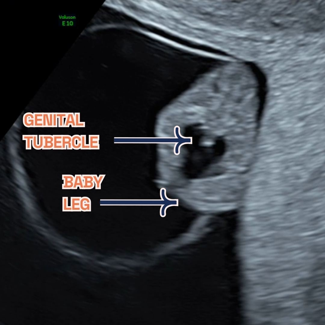
By week 9, the genital tubercle is visible in all embryos, signifying a key phase in gender development. Visible via 2D or 3D ultrasound, its clarity improves with transvaginal scans. This method provides high-resolution images, offering a clear glimpse of early baby movements. While each pregnancy is unique, ultrasound scans follow a similar process. As time goes on, the genital tubercle evolves into either a penis or a clitoris, marking the baby’s gender.

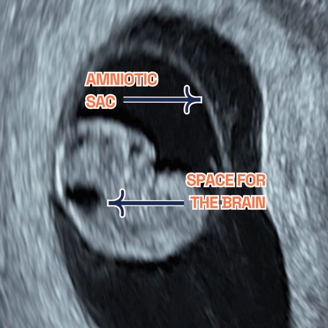
  • Image showing what would an ultrasound show at 8 weeks of pregnancy. It shows an Ultrasound image by London Pregnancy Clinic that shows the gestational sac, fetal pole, amniotic sac and yolk sac - for educational purpose.

    Gestational sac, fetal pole and amniotic sac at 8 weeks of pregnancy as seen on ultrasound scan.

  • Gestational sac, fetal leg and Genital Tubercle at 9 weeks of pregnancy as seen on ultrasound scan.

    Gestational sac, fetal legs and Genital Tubercle at 9 weeks of pregnancy as seen on ultrasound scan.

Thinking ahead: Week 10?

Around 10 weeks into pregnancy, some women might had their initial ultrasound scan already. This scan helps confirm the due date by measuring the baby’s size and checks for multiple births or genetic conditions like Down’s syndrome risk. It’s crucial to keep regular prenatal check-ups and share any concerns with healthcare professionals for proper advice during pregnancy. Discuss the option of a NIPT with your doctor. Available from week 10, this test screens for chromosomal abnormalities and can reveal the baby’s sex.

You may be considering our innovative 10 Week Scan, it’s important to note that this is distinct from the usual viability or dating scans you might be familiar with. Specifically designed for 10-11 weeks of pregnancy, this scan acts as the earliest anomaly check, providing parents with early reassurance by screening for significant physical abnormalities. It marks the first opportunity for a structural examination of the baby, at the earliest stage possible. This timing also makes it the optimal period for non-invasive prenatal testing (NIPT), leveraging our blend of professional expertise and the latest in ultrasound technology to deliver results with unmatched clarity. Compared to the standard NHS 12 week scan, our 10-week scan offers a more comprehensive checklist, ensuring nothing is overlooked. 

Remember, NIPT indicates the likelihood of certain conditions, unlike amniocentesis or chorionic villus sampling (CVS), which provide definitive results but are invasive. Always check if your insurance covers NIPT. For those over 35 or with a family history of genetic conditions, consider NIPT, CVS (done between 10 and 13 weeks), and amniocentesis (usually between weeks 15 to 20). The likelihood of a baby having a chromosomal condition, such as aneuploidy, rises with maternal age, notably after 35 years, termed “advanced maternal age.”

Week 9: Your body?

Alongside fetal developments, maternal changes may be observed during the ultrasound. Increased blood volume and fluid retention can lead to frequent urination. Hormonal fluctuations may cause symptoms like headaches, low blood pressure, constipation, and fatigue. These changes reflect the body’s adaptation to support the growing fetus during pregnancy.

Keep hydrated by drinking plenty of water, aiming to drink more in the morning to avoid nighttime trips to the loo. At 9 weeks, you might struggle to fasten your jeans as your uterus expands, doubling in size to accommodate your growing baby. You may even start to show a little. As the weeks progress, your uterus will grow beyond your pelvis. Gaining weight now is not just normal—it’s advisable. Your ideal weight gain during pregnancy depends on your pre-pregnancy BMI. Generally, with a normal BMI, you’d be expected to gain 25 to 35 pounds throughout, with one to five pounds in the first trimester.

However, morning sickness and food aversions may lead to weight loss instead of gain at this stage. Discuss any weight concerns, gain or loss, especially if changes are sudden or severe, with your obstetrician or midwife. Minor weight loss is often seen as acceptable early on. As your appetite returns, there’ll be chances to get your weight gain on track. Severe nausea may require medical intervention. Many under-report nausea and vomiting, but numerous safe Over-The-Counter and prescription options can manage symptoms. 

Week 9 of Pregnancy FAQs


  • what happens during week 9 of pregnancy?

    At 9 weeks pregnant, your body and baby undergo significant changes. Not everyone experiences the same symptoms, ranging from hair thickening—due to reduced shedding and possibly healthier lifestyles including prenatal vitamins—to bloating, caused by increased progesterone slowing digestion.

    An early scan reveals your baby’s rapid growth, from forming hands and feet to facial features developing, and even early movements. The scan also shows the brain’s development, the steady heartbeat indicating a strong cardiovascular system, and the placenta’s vital role in providing nutrients. The genital tubercle’s visibility marks the beginning of gender development, becoming clearer with transvaginal scans.

  • spotting during week 9 of pregnancy?

    Spotting in week 9 of pregnancy is commonly seen and typically isn’t a cause for concern. Light bleeding or spotting can be normal in the first trimester. Nonetheless, consulting your midwife or healthcare provider for a check-up is always wise to dismiss any potential issues.

    Usually, spotting isn’t alarming, but monitoring the situation and getting medical advice if the bleeding gets heavy or continuous is vital. Keeping in contact with your healthcare provider during your pregnancy is crucial to quickly address concerns and safeguard the health of you and your baby.

    Every pregnancy is distinct, and although spotting can be standard, consulting your healthcare provider for tailored advice and reassurance is always recommended.

  • why is week 9 of pregnancy the worst?

    Week 9 of pregnancy might bring challenges due to physical and hormonal changes. Your breasts may enlarge and your waist could thicken as your uterus grows. High levels of pregnancy hormones may cause discomfort and morning sickness. These symptoms are often temporary and improve by the second trimester. Nausea and vomiting, known as “morning sickness,” can happen any time. This is often due to the rise in human chorionic gonadotropin (hCG), peaking around week 10. So, the most intense nausea might occur between now and next week. Additionally, increased progesterone can make your breasts larger or swollen.

  • Common symptoms during nine weeks pregnant

    Feeling early flutters at 8 weeks? Expect:

    • Fatigue: Your body is working hard, so tiredness is typical. Make rest a priority.
    • Frequent urination: Your expanding uterus presses on the bladder, meaning more loo visits.
    • Morning sickness: Nausea and vomiting might strike any time. Keep hydrated and seek relief methods.
    • Enhanced senses: You may experience metallic tastes or strong smells. It’s all part of the journey!
    • Mood swings: Hormonal changes may affect your emotions. Treat yourself gently.
    • Sore breasts: Hormones are prepping for breastfeeding, causing discomfort.
    • Bloating and cramping: These are signs of your growing uterus.
    • Light spotting: It’s rare but can occur. Contact your doctor if worried.
    • Other signs: Thicker hair, skin changes, or milky discharge are normal during pregnancy.

    Each pregnancy differs. Some women get many symptoms; others have few. Always consult your healthcare provider with concerns. Consider a private midwife consultation for worries.

  • Can I do NIPT at 9 weeks of pregnancy?

    Non-invasive prenatal testing (NIPT)

    can start at 9 weeks into pregnancy. It’s a safe, accurate way to screen for chromosomal abnormalities like Down’s, Edwards’, and Patau’s syndromes. By analysing the mother’s blood for cell-free DNA, NIPT gauges the fetus’s risk for specific genetic conditions without miscarriage risks, ensuring early detection and reassurance. We advise waiting until week 10 for NIPT, allowing it to coincide with our 10-week scan for optimal results.

    NIPT stands out for its precise detection of chromosomal disorders, showing high sensitivity for conditions such as Down’s syndrome. Early testing allows for informed decisions and tailored prenatal care. Discussing NIPT with your healthcare provider is crucial to see if it suits your needs and to understand test implications. 

    Benefits of NIPT include:

    • Non-invasive with no risk to the baby.
    • High accuracy for peace of mind.
    • Early detection for proactive decision-making.
    • Optional sex determination for early family planning.

    Remember: NIPT is a screening test, not a definitive diagnosis. Consult your healthcare provider for further information and personalised guidance. We at LPC offer multiple NIPTs (PrenatalSafe, Panorama, Unity, SMART TEST, Illumina, Harmony) and out specialists will help you choose the best option for you!


Final Thoughts

Week 9 is a time of remarkable progress, and London Pregnancy Clinic is here to guide and support you through every step. With each visit, you’ll gain deeper insights into your baby’s development, reinforcing the bond you share. Embrace this journey with confidence, knowing we’re by your side.

Stay informed and excited about what each new week will reveal about your baby’s growth on our blog.


Latest Stories

Continue reading

Week 8 of Pregnancy

Graphic of Week 8 of pregnancy. An illustration of a baby at week 8 in the gestational sac.

Week 8 of your Pregnancy

Discovering your baby’s progress since week 7

  • Published

  • Last Modified

Tags

Embark on the captivating journey from Week 7 to Week 8 of your pregnancy with the London Pregnancy Clinic. Witness the awe-inspiring progress of your baby’s development with our private early pregnancy scans. Week 8 is a milestone where your baby begins to move and grow at an astonishing rate, from tiny limb movements to the development of internal organs. Embrace the changes and find reassurance in observing your baby’s advancement.

As you step into Week 8 of your pregnancy, you’re entering a crucial phase of development. Your baby, now rapidly evolving, is preparing for the next significant steps in prenatal growth. At London Pregnancy Clinic, we share in your excitement and curiosity during this time. This week is often when expectant mothers, seek reassurance and connection to their growing baby through our private early pregnancy scans.

Key Concepts: fetal development

Movement and Growth: By Week 8, your baby, now the size of a raspberry (around 15mm), begins to move, although you might not feel it yet. These early movements are a sign of muscle development and coordination. The ultrasound may capture some of these movements, providing a heartwarming glimpse for expectant parents.

Organ and Brain Development: Major organs, muscles, and nerves start functioning, with the brain forming primitive connections between nerve cells, laying down the first neural pathways. This week also allows us to measure brain waves for the first time, a testament to the burgeoning complexity of your baby’s development.

Physical Features: The facial features of your baby become more defined, with the formation of the upper lip, the tip of the nose, and the eyelids. The limbs extend, and fingers and toes start to emerge from their webbed structure, marking a significant step towards their final shape.

Pregnancy Checklist at 8 Weeks

  • Book and attend your private early pregnancy scan: Capturing clear images during the 6-9 week Viability Scan is crucial for assessing your baby’s development and health. These dating Scans are pivotal, offering crystal-clear imagery that plays a crucial role in monitoring your baby’s development and ensuring their health. Beyond the clinical benefits, these scans foster a profound emotional bond, providing expectant parents with a reassuring glimpse into their baby’s growth and vitality. Our transvaginal scans employ advanced 3D/4D ultrasound technology, enabling us to capture high-resolution images of your baby’s earliest movements, bringing you closer to the miracle of life unfolding within you.
  • Focus on your health and wellness: Your baby’s development relies on your well-being. Ensure you maintain a balanced diet, get regular, gentle exercise, and follow your healthcare provider’s advice.
  • Communication with healthcare professionals: Maintaining open and consistent communication with your healthcare professionals is paramount during this transformative period. Whether navigating the physical changes sweeping through your body or adjusting to the hormonal shifts, having a trusted medical advisor by your side can make all the difference. They are your primary source of support, guidance, and reassurance, dedicated to ensuring your pregnancy journey is as smooth and informed as possible.
  • Booking Appointment: If you haven’t yet scheduled your first antenatal appointment, now is the time to consider this essential step. The NHS offers comprehensive antenatal care starting with the booking appointment, which is a cornerstone of your prenatal care. Alternatively, our private midwife services at London Pregnancy Clinic provide a personalised care experience, ensuring you receive tailored advice and support that resonates with your unique pregnancy needs. This initial consultation is an invaluable opportunity to ask questions, express concerns, and set the foundation for a healthy pregnancy.

Developmental Milestones: Week 8

Embryonic Growth: The rapid growth, your little one measures approximately 15 to 20 mm in length, also known as the crown to rump or CRL. The embryonic form transforms, with fingers and toes delineating further, shedding their webbed guise for a more distinct appearance. 

Gestational and Yolk Sac Development: At 8 weeks the gestational sac typically ranges from 27 to 33 mm. The yolk sac is about 3.3 mm. The nurturing environment of your baby, the gestational sac, demonstrates healthy growth, enveloping your baby in a protective embrace. The yolk sac, though still visible, will soon pass the baton to the placenta, becoming the primary source of nourishment for your developing baby.

Heart Development: Fetal heart rate slightly increases to an average of 140-170 beats per minute (BPM) and will gradually stabilise around 12 weeks.

Other Development: By the week’s close, the blueprint for all essential organs and body systems is firmly laid out. The foundations for your baby’s gender are being set, though the grand revelation remains a few weeks away (too soon to see on ultrasound). Meanwhile, the eyes begin their journey with the retinas starting to form.

Umbilical cord: Formed by week 7, this vital cord acts as a highway, connecting your baby to the placenta. It facilitates a critical exchange: oxygen and nutrients flow in via the umbilical vein, while waste products exit through the umbilical arteries. At this stage, the cord measures approximately 50-60cm, possibly exhibiting up to 40 helical turns for added strength and flexibility.

What is seen on the Ultrasound: Week 8?

The heartbeat is now stronger and more rhythmic, it serves as a profound indicator of your baby’s health and vitality. For expectant parents of twins, this week may unveil the presence of two heartbeats, doubling the joy and anticipation. The ultrasound meticulously charts the development of each embryo, ensuring both are progressing beautifully on their journey.

A subtle opening in the baby’s head marks the space where the brain will continue to grow and flourish. Far from alarming, this natural process is a testament to the body’s remarkable capacity to adapt and prepare for the months ahead. The ultrasound captures this phase with care, offering a glimpse into the brain’s early stages of development in a way that’s both reassuring and awe-inspiring. 

Another fascinating aspect of this scan is the appearance of two parallel lines, a preliminary indication of the fetal spine’s formation. This early sign of structural development underscores the incredible precision with which life unfolds. Both the umbilical cord and amniotic sac, vital to your baby’s development, will be seen during the scan. The umbilical cord, a lifeline to nourishment and oxygen, and the amniotic sac, a protective cocoon, are both visible. Together, they create an environment where your baby can grow, move, and thrive – which you may witness during the scan.

To ensure the most accurate observation, we may utilise transvaginal ultrasounds during these early stages. This method allows us to obtain high-resolution images and a clearer view of your baby’s early movements, compared to the traditional abdominal ultrasound. All pregnancies are unique but what happens during the ultrasound scan is similar for everyone.

  • Image showing what would an ultrasound show at 7 weeks of pregnancy. It shows an Ultrasound image by London Pregnancy Clinic that shows the gestational sac, fetal pole and yolk sac - for educational purpose.

    Gestational sac, fetal pole and yolk sac at 7 weeks of pregnancy as seen on ultrasound scan.

  • Image showing what would an ultrasound show at 8 weeks of pregnancy. It shows an Ultrasound image by London Pregnancy Clinic that shows the gestational sac, fetal pole, amniotic sac and yolk sac - for educational purpose.

    Gestational sac, fetal pole and amniotic sac at 8 weeks of pregnancy as seen on ultrasound scan.

Thinking ahead: Week 9?

Looking forward to Week 9, anticipate witnessing continued growth and development during your next scan. Maintain a healthy lifestyle and prepare for your upcoming prenatal appointments. Consider discussing prenatal screening options (like NIPT, which we offer complimentary counselling for during our Viability Scan) and dietary adjustments with your healthcare provider to support this vital growth phase.

Morning sickness may persist during Week 8, but it’s a common and typically harmless part of pregnancy. It’s often a result of hormonal changes and is expected to improve as you approach the second trimester. For severe cases, seek professional advice for management strategies.

Week 8 of Pregnancy FAQs


  • 8 week pregnancy is how many months?

    At 8 weeks pregnant, you’re right around 2 months along, though doctors usually track pregnancy progress by week for more precise development details. This means you have roughly 7 months until your amazing arrival! Remember, this is just an estimate, and your little one might choose to come a bit earlier or later. Exciting times ahead!

  • what’s happening during week 8 of pregnancy?

    At 8 weeks pregnant, your little one is making incredible progress! Here’s the summary:

    • Grows rapidly to 15-20mm (crown-to-rump).
    • Fingers and toes become more distinct, losing their webbed appearance.
    • Heart rate increases to 140-170 BPM and will stabilize soon.
    • Blueprint for all organs and body systems is established.
    • Gender starts to develop, but still too early to reveal on ultrasound.
    • Eyes begin to form with developing retinas.
    • Gestational sac grows to 27-33mm, protecting the baby.
    • Yolk sac still visible but becoming less important as the placenta takes over as the main source of nourishment.
    • Umbilical cord (formed in week 7) transports oxygen and nutrients to the baby and removes waste. It’s about 50-60cm long and might have twists for stability.
  • week 8 of pregnancy and no morning sickness?

    At 8 weeks pregnant, you might be surprised to be experiencing no morning sickness, especially if it’s a common concern among expectant parents. Rest assured, the absence of nausea and vomiting during pregnancy does not necessarily indicate any underlying issues.

    Here’s what you need to know:

    • Individual Response: The experience of pregnancy symptoms like morning sickness varies greatly among individuals. While it affects 70-80% of pregnant people, the remaining 20-30% may report minimal to no symptoms.
    • Fluctuating Symptoms: Even within the same individual, symptom intensity can fluctuate throughout pregnancy. While you might feel good now, mild nausea could still develop later.
    • Spectrum of Severity: “Morning sickness” encompasses a range of experiences, from occasional mild nausea to frequent vomiting. You might be falling on the less intense end of the spectrum.

    Therefore, the absence of morning sickness at 8 weeks is not a cause for concern. However, if you have any questions or worries about your pregnancy, or experience other concerning symptoms, always consult your healthcare provider for personalised guidance and reassurance.

  • Common symptoms during eight weeks pregnant

    Feeling the early flutters? Here’s what to expect at 8 weeks:

    • Fatigue: Your body’s working overtime, so exhaustion is common. Prioritise rest and listen to your needs.
    • Frequent urination: The growing uterus puts pressure on your bladder, leading to more bathroom trips.
    • Morning sickness (and maybe not just in the mornings): Nausea, vomiting, and food aversions are a rollercoaster for many. Stay hydrated and find remedies that work for you.
    • A symphony of sensations: From metallic tastes to heightened smells, your senses might be on overdrive. Embrace the unique experience!
    • Mood swings: The hormonal dance can stir up emotions. Be kind to yourself and seek support if needed.
    • Sore breasts: This is all thanks to hormonal changes preparing for milk production.
    • Bloating and cramping: Your uterus is expanding, which can cause these temporary discomforts.
    • Light spotting: While uncommon, it can happen. If concerned, consult your doctor.
    • Other changes: You might notice thicker hair, skin pigmentation changes, or a milky discharge – all normal pregnancy variations.

    Every pregnancy is unique. Some women experience all these symptoms, while others have none. If you have any concerns or questions, don’t hesitate to talk to your healthcare provider.

    Note: The information provided in this blog is intended for educational purposes only and should not be considered as a substitute for professional medical advice, diagnosis, or treatment. 

  • I am bleeding when I’m 8 weeks pregnant – what do i do?

    Bleeding during pregnancy, even at 8 weeks, requires immediate medical attention. While occasional light spotting can be harmless, it’s crucial to seek professional guidance right away, as bleeding can also be a sign of a more serious issue. Early pregnancy can experience bleeding due to various factors, including implantation, cervical changes, miscarriage, or ectopic pregnancy.

    To ensure the well-being of both you and your baby, contact your midwife or GP without delay, especially if the bleeding is persistent or accompanied by symptoms like severe pain or dizziness. They may conduct an examination, ultrasound, or blood tests to determine the cause and provide appropriate care.

    Remember, prioritising your health and seeking prompt medical attention is vital when experiencing any unexpected bleeding during pregnancy.

    Note: This information is for educational purposes only and should not be considered medical advice. Please consult with your healthcare provider for any questions or concerns you may have regarding your pregnancy, especially if you experience any bleeding.

  • 8 weeks pregnant. Twins?

    At 8 weeks, twin miracles are developing! Their tiny bodies, about 15m cm long and 1 gram each, showcase rapid progress. Limbs and kidneys form and human features become distinct. Beating hearts, now divided into chambers, race at 150 BPM.

    Ultrasound magic at 8 weeks might reveal twin gestational sacs, fetal poles, and even fluttering heartbeats, confirming your double joy. However, it’s vital to be aware of vanishing twin syndrome, where one twin stops developing in early pregnancy. While symptoms like morning sickness and fatigue could be more intense with twins, discuss any concerns with your healthcare provider. They can guide you through this unique pregnancy journey and ensure optimal care for you and your remaining little miracle.

  • Week 8 pregnancy bump…

    At 8 weeks pregnant, don’t expect a visible bump just yet! Your little one is still tiny! While you might feel bloated or have slightly swollen breasts due to hormonal changes, a true baby bump typically appears closer to 12-16 weeks, when your uterus grows enough to peek out of your pelvis.

    Here’s what’s happening instead:

    • Rapid internal growth: Your baby is forming all their essential organs and systems at lightning speed!
    • Uterine expansion: Your uterus starts expanding to make room for your growing baby, but it’s still tucked inside your pelvis.
    • Bloating: Bloating and gas due to hormonal changes are common early pregnancy symptoms, causing a temporary “puffy” feeling.

    So, relax and enjoy this early stage of pregnancy! Remember, every woman’s body experiences pregnancy differently, and a visible bump can appear earlier or later depending on various factors.

  • NIPT? what’s that?

    Non-invasive prenatal testing (NIPT) is a groundbreaking screening tool offering peace of mind during pregnancy. This simple blood test examines cell-free fetal DNA in your blood to assess the risk of specific chromosomal abnormalities in your baby:

    Down’s syndrome (Trisomy 21): Detected with >99% accuracy.
    Edward’s syndrome (Trisomy 18): Detected with 97.4% accuracy.
    Patau’s syndrome (Trisomy 13): Detected with 93.8% accuracy.

    These figures represent detection rates reported by Harmony NIPT. Additionally, NIPT boasts a remarkable <0.1% false positive rate for all three trisomies, calculated at a 1/100 risk cutoff.

    And there’s more! NIPT can also determine your baby’s sex with >99% accuracy, offering an early glimpse into your growing family.

    Key benefits of NIPT:

    • Non-invasive: No needles or risks to your baby.
      Highly accurate: Provides valuable information about your baby’s health.
    • Early detection: Offers time for informed decision-making if needed.
    • Optional sex determination: Satisfies your curiosity early on.

    Remember: NIPT is a screening test, not a definitive diagnosis. Consult your healthcare provider for further information and personalised guidance. We at LPC offer multiple NIPTs (PrenatalSafe, Panorama, Unity, SMART TEST, Illumina, Harmony) and out specialists will help you choose the best option for you!


Conclusion

At Week 8, while some challenges may arise, such as the potential for hyperemesis gravidarum or the need for reassurance regarding pregnancy progression, London Pregnancy Clinic is here to support you. Our expertise and advanced scanning technology provide the care and answers you need during this transformative time.

Stay tuned for more updates as you progress through this incredible journey, and remember, our team is always here to support you every step of the way.


Latest Stories

Continue reading

Week 7 of Pregnancy

Graphic of Week 7 of pregnancy. An illustration of a baby at week 7 in the gestational sac.

Week 7 of your Pregnancy

Discovering your baby’s progress since week 6

  • Published

  • Last Modified

Tags

Embark on the transformative journey from Week 6 to Week 7 of your pregnancy with London Pregnancy Clinic. Experience the wonder of your baby’s growth with our private early pregnancy scans. Week 7 brings rapid development, from distinct heartbeats to the formation of facial features. Embrace the new changes and the reassurance of seeing your baby’s progress.

Stepping into Week 7 of your pregnancy, you’re embarking on a remarkable phase. Your baby, swiftly growing and developing, is now ready for the next stage of prenatal monitoring. At London Pregnancy Clinic, we understand the excitement and curiosity that comes with this week. It’s a time when many expectant mothers like Sarah and Emily seek reassurance through our private early pregnancy scans.

Key Concepts: fetal development

Baby’s Fast Heart Beat: During week 7, your baby’s heart rate accelerates, typically beating between 120 and 160 times per minute. This rapid heartbeat is a healthy sign of your baby’s growth and development. It’s an exhilarating moment for many parents, often heard for the first time during a viability scan.

Facial Features: By Week 7, your baby, roughly the size of a blueberry, shows significant development. The viability scan often reveals more distinct features and movements. The embryo’s facial features start taking shape, with the brain developing at a remarkable pace.

Rapid Development: 7 weeks marks the start of a huge growth spurt. During this period (between 7 and 20 weeks) your baby’s body parts will grow rapidly and organs such as the heart and brain will develop and increase in complexity. The basic structures of all major body parts and organs are formed during this time, with much of the growth being focused on the brain. Your baby’s brain cells are being generated at a rate of 100 per minute! The first blood vessels of the circulatory system appear. The appendix and pancreas are now evident, and inside the mouth, tooth buds, a tongue and the palate are all developing.

Pregnancy Checklist at 7 Weeks

  • Book your private early pregnancy scan: Clear 7-week scan images can provide valuable information about your baby’s development and well-being. Ultrasound scans, especially the Viability Scan, offer emotional moments for expectant parents, providing a tangible connection to their growing baby and reassurance about their well-being. Despite natural concerns or anxieties, these routine and safe procedures offer valuable insights into the baby’s development.
  • Monitor your health and well-being: As your baby continues to grow rapidly, take care of yourself by maintaining a healthy lifestyle, eating nutritious foods, and staying active as recommended by your healthcare provider.
  • Keep an eye on your nutrition: Keep focusing on foods supporting fetal development and ensuring you get essential vitamins and minerals.
  • You’re not alone, talk to professionals: During the early stages of pregnancy, it’s crucial to communicate with your healthcare provider as your hormones and body begin to change. They can offer guidance, support, and reassurance for any questions or concerns. Your provider has the expertise to address any issues or uncertainties that may arise, so don’t hesitate to reach out to them whenever needed.

Developmental Milestones: Week 7

Embryonic Growth: The embryo now measures between 9mm to 10mm long (from crown to rump – CRL) and is 10,000 times bigger than it was at conception. Your baby’s fingers and toes are now formed, although not separated, giving them a paddle-like appearance. The embryo shows flurries of rapid movements at this stage. The liver is now producing red blood cells, and will continue to do so until bone marrow is formed which then takes over the production of red blood cells.

Gestational and Yolk Sac Development: At 7 weeks the gestational sac typically ranges from 18 to 25 mm. The yolk sac is about 3 mm. Both continue to grow steadily. The yolk sac provides nutrients and aids in blood cell formation until the placenta takes over this role. In Week 7, the gestational and yolk sacs play a critical role in your baby’s development. The gestational sac envelops the embryo, but its presence doesn’t guarantee a normal, healthy pregnancy. An anembryonic pregnancy, or blighted ovum, may occur, where the gestational sac lacks an embryo. This can be caused by various factors. If they are significantly smaller, it may indicate a potential first-trimester pregnancy loss. Seek guidance from early pregnancy specialists and obstetric professionals for further evaluation and support.

Heart Development: Between weeks 6 and 7 of pregnancy, the baby’s heart rate speeds up, usually ranging between 120 and 160 beats per minute.

Baby Features Development: By the end of week 6, the ears are continuing to form while the eyes are now covered with the beginnings of what will later become eyelids. Now at week 7, your baby’s fingers and toes are formed, although not separated, giving them a paddle-like appearance. 

What is seen on the Ultrasound: Week 7?

As you enter Week 7 of your pregnancy, the ultrasound offers a more intricate view of your baby’s development, building upon the observations from Week 6. This scan is a window into a world of rapid growth and transformation taking place within you. During the 7-week ultrasound, expect to see enhanced and more distinct images. The heartbeat, which was a delicate flicker last week, is now more pronounced and rhythmic, beating confidently as a testament to your baby’s vitality. 

The gestational sac, your baby’s first nurturing environment, continues to grow, signifying healthy progression. Inside this protective sac, the yolk sac remains visible, providing essential nourishment. It’s a critical component until the placenta fully develops and takes over the role of supporting your baby.

The embryo itself is undergoing remarkable changes. Now resembling a tiny being with a more defined head and a tail-like structure, the embryo’s curved form is more noticeable. This is the period when major organs start forming. The healthcare provider may measure the embryo from head to tail, known as the CRL. Alongside, the size of the gestational sac is also measured, providing further information about the pregnancy’s progression.

This week’s ultrasound is pivotal for confirming the pregnancy’s viability, as it checks for the presence of a heartbeat and helps rule out any complications. Depending on the clarity needed, the ultrasound may be performed trans-abdominally or transvaginally, with the latter often providing a clearer view of these early developmental stages. Each pregnancy is unique, and these developmental milestones may vary slightly from one individual to another but what to expect during the ultrasound scan will remain the same.

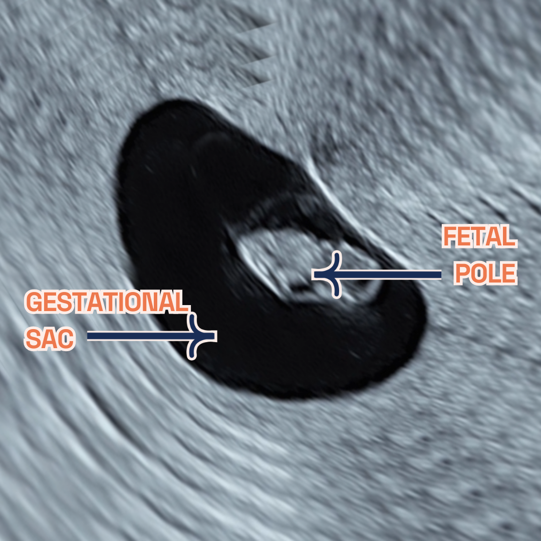
  • Image showing what would an ultrasound show at 6 weeks of pregnancy. You can see the fetal pole, gestational sac and yolk sac. It shows an Ultrasound image by London Pregnancy Clinic that shows the gestational sac and how small it is - for educational purpose.

    Gestational sac, fetal pole and yolk sac at 6 weeks of pregnancy as seen on ultrasound scan.

  • Image showing what would an ultrasound show at 7 weeks of pregnancy. It shows an Ultrasound image by London Pregnancy Clinic that shows the gestational sac, fetal pole and yolk sac - for educational purpose.

    Gestational sac, fetal pole and yolk sac at 7 weeks of pregnancy as seen on ultrasound scan.

Thinking ahead: Week 8?

As you approach Week 8, expect further growth and development. Your next scan will provide more details about your baby’s progress. Keep following health guidelines and get ready for your next prenatal appointment. To support this critical growth phase, discuss upcoming screenings (we offer free counselling during our Viability Scan for NIPT) and nutritional adjustments with your healthcare provider. Approximately 75% of pregnant women experience morning sickness, which is a common symptom during week 7 of pregnancy. Rest assured, it’s a normal part of the journey. Despite its name, it can happen at any time of the day and is likely caused by hormonal changes and uterine muscle stretching. Nausea and vomiting usually improve between weeks 12 to 14 of pregnancy. Seek medical advice for severe morning sickness and remember, relief is on the way in a few weeks! Hang in there!

Conclusion

Week 7 of Pregnancy FAQs


  • How does 7 weeks pregnant look like?

    At 7 weeks pregnant, your tiny miracle experiences remarkable growth!

    • Baby development: This stage marks rapid progress. Your little one, roughly the size of a grape (around 9-10mm long), showcases significant development. The brain, crucial for future cognitive function, grows quickly, leading to a prominent forehead. Meanwhile, eyes and ears continue their formation, laying the groundwork for sight and hearing. Tiny limb buds, precursors to arms and legs, begin to transform into cartilage, the foundation for future bones. Nerve cells multiply rapidly, supporting your baby’s incredible growth spurt.
    • Maternal experiences: As your baby thrives, your body adapts. The womb expands to accommodate your growing miracle, reaching around the size of a lemon. Common symptoms like fatigue, tender breasts, frequent urination, and morning sickness might arise, though remember, that every pregnancy is unique, and your experiences may differ.

    Disclaimer: Remember, this information is for general knowledge only and should not be considered a substitute for professional medical advice. Always consult your healthcare provider for personalised guidance and to address any concerns you may have.

  • What to expect when 7 weeks pregnant?

    As your baby flourishes, you may notice personal changes as well:

    • Morning Sickness: A common symptom, though it doesn’t affect everyone.
    • Fatigue: It’s normal to feel unusually tired as your body nurtures your growing baby.
    • Sore Breasts: Hormonal changes may cause tenderness or swelling.
    • Food Preferences: You might find your taste preferences changing.
    • Enhanced Sense of Smell: Scents may become more intense.
    • Mood Fluctuations: Hormones can lead to emotional variability.

    Additional experiences some women report include:

    • Metallic Taste: An odd but temporary sensation.
    • Headaches: Ensure you’re well-hydrated and consult a doctor if headaches are frequent.
    • Light Spotting: While it may be alarming, it’s not always a sign of trouble. Seek advice from your healthcare provider for reassurance.
    • Cramps: These can resemble period pains but are generally less severe.

    Advice to Remember:

    • Stay Hydrated: Drinking water is crucial for your changing body.
    • Maintain a Healthy Diet: A balanced diet benefits both you and your baby.
    • Listen to Your Body: Rest as necessary and focus on self-care.
    • Seek Support: For any concerns, professional guidance from your healthcare provider is invaluable.

    Each pregnancy journey is distinct. Embrace this special time and the transformations it brings.

  • Can you feel movement seven weeks of pregnancy?

    At 7 weeks into pregnancy, feeling your baby’s movements is usually too soon. Typically, the initial signs of fetal movement, or quickening, are experienced between 13-25 weeks, with some noticing these sensations as early as 13-16 weeks. Initially described as a flutter, these movements can be subtle, often mistaken for gas or similar sensations. The timing of when these movements are felt can be influenced by various factors, including the placenta’s position, the volume of amniotic fluid, and the mother’s body mass index. However, at week 8 of pregnancy, the baby starts moving inside the womb and that can be seen during an ultrasound scan.

  • I am bleeding when I’m 7 weeks pregnant – what do i do?

    Prompt medical attention is crucial for bleeding at 7 weeks of pregnancy. Light spotting may not always be concerning, but professional consultation is essential to rule out serious conditions such as miscarriage or ectopic pregnancy. Immediately contact your midwife or GP, especially if accompanied by severe pain or dizziness, for assessment and appropriate care.

    Note: This is educational content and not medical advice. Consult your healthcare provider for any pregnancy-related concerns, especially bleeding.

  • I am having twins during my 7 weeks viability scan

    Discovering you’re expecting twins at your 7-week viability scan is a possibility, thanks to the precision of modern ultrasound technology. Particularly, a transvaginal ultrasound can effectively identify twins as early as 6 weeks into pregnancy. This type of scan can reveal the presence of multiple gestational and yolk sacs, fetal poles, and even distinct heartbeats, all indicative of a twin pregnancy. Notably, during the 7-9 week period, transvaginal ultrasounds are highly reliable for assessing the specifics of twin pregnancies, such as chorionicity and amnionicity.


Conclusion

Week 7 is a period of significant progress in your pregnancy. London Pregnancy Clinic is here to support you with expert care and detailed scans. Remember, every pregnancy is unique, so embrace your journey with confidence and excitement. Stay tuned for our next update as you continue this incredible journey.

Stay tuned on what your baby’s week-by-week growth will look like on our blog.


Latest Stories

Continue reading

Week 6 of Pregnancy

Minimalist flat design illustration of a 6-week fetus resembling a small tadpole next to a large yolk sac, representing an ultrasound image for a private early pregnancy scan at the specialist London Pregnancy Clinic, capturing the essence of a 6 weeks early pregnant ultrasound scan.

Week 6 of your Pregnancy

Discovering your baby’s progress since week 5

  • Published

  • Last Modified

Tags

Having progressed from the foundational moments of Week 5, Week 6 ushers in a new chapter of pregnancy. It’s a time when your little one, just past the size of an apple seed, shows the first signs of a heartbeat, symbolising vibrant growth and transformation. This period marks an exciting phase of development, building upon the crucial milestones of Week 5.

Welcome to Week 6 of your pregnancy journey, an exhilarating time filled with significant milestones. This week often marks the first opportunity for mothers-to-be to witness the wonders of their growing baby through early scans. As the heartbeat begins and the baby continues to develop, understanding the changes and knowing what to expect can provide comfort and excitement during this special time.

Key Concepts: fetal heartbeat

The First Beat: Around 6 weeks, your baby’s heart begins to beat with a rhythm that will continue to guide their growth throughout the pregnancy. This is a critical milestone in cardiac development, indicating healthy progression.

Observing the Heartbeat: During a 6 week ultrasound, often called a Viability/Dating Scan, you might be able to see the fetal heartbeat for the first time. It’s a profound moment for many parents, symbolising the new life growing inside.

Understanding the Rate: A fetal heartbeat at 6 weeks can range between 90-110 beats per minute and will increase in the coming weeks. This rapid rate supports the fetus’s growing need for oxygen and nutrients as development accelerates. The heart rate is a vital indicator of fetal health. A strong and clear heartbeat offers reassurance of the pregnancy’s viability and normal development.

Pregnancy Checklist at 6 Weeks

  • If you haven’t already, now is the time to schedule your first private early pregnancy scan. This will confirm the pregnancy’s viability and provide valuable information about the baby’s development. You can read more on what to expect during your ultrasound scan here.
  • Continue focusing on nutrition, especially foods rich in folic acid, iron, and calcium. Stay hydrated and get plenty of rest to support your body’s increased needs.
  • Be aware of common symptoms such as morning sickness, fatigue, and breast tenderness. Find strategies that help manage these symptoms, whether it’s through dietary changes, rest, or gentle exercise.
  • As your body continues to change, consider comfortable clothing options and start planning for future needs. Also, begin to think about discussing your pregnancy with your employer to understand any potential work adjustments or benefits. Sharing your news will come naturally when you’re ready, but taking care of yourself and planning ahead? Now that’s just blooming smart!

Developmental Milestones: Week 6

As you enter Week 6, your baby is rapidly growing and developing. Here are some of the key milestones:

Embryonic Growth: The baby, now about the size of a sweet pea, is growing quickly. The length may be around a quarter of an inch or 3-4mm. At this stage of pregnancy, the baby will continue to grow at a pace of 1mm a day. This is a critical period for organ formation. The brain, heart, and other vital organs begin to take shape and grow.

Yolk Sac Development: At 6 weeks of pregnancy, the yolk sac plays a pivotal role in the development of your baby. During this time, it’s normal for the yolk sac to be slightly larger than the fetus. Typically, it continues to grow, usually in a linear fashion until around 10 weeks of gestation. It’s responsible for providing nutrients to the developing fetus and assisting in blood cell formation until the placenta is fully developed and ready to take over this role.

Heart Development: One of the most monumental moments in early pregnancy occurs — the detection of the fetal heartbeat. While it might still be early, and not all scans will reveal a heartbeat at this stage, many parents can witness the rhythmic flickering of their baby’s tiny heart on the ultrasound screen.

Baby Features Development: Tiny indentations where the eyes will develop and small buds that will grow into ears start forming, laying the foundation for your baby’s face. Likewise, limb buds begin to emerge, signaling the future development of arms and legs. These developments are subtle yet signify a period of rapid and complex growth as the basic structures and organ systems start forming.

What is seen on the Ultrasound: Week 6?

As you progress into Week 6 of your pregnancy, the ultrasound experience evolves, bringing new developments into view. Building upon the foundational observations of Week 5, the Week 6 ultrasound typically offers a more detailed glimpse into your baby’s development.

The gestational sac continues to house and protect your developing baby and its increased size from the previous week is a positive indication of ongoing growth and development. Moreover, the yolk sac will still be visible and may have increased in size, mirroring the embryo’s growth. It continues to perform its vital role until the placenta is ready to take over.

One of the most significant and emotional moments that happens when you are doing the 6 weeks pregnant ultrasound is the detection of the heartbeat. Unlike Week 5, where the embryo and its heartbeat are usually not yet visible, the viability scan often provides the first visual confirmation of the baby’s heartbeat. This is seen as a rhythmic flickering on the ultrasound screen.

In addition to the heartbeat, you might also start to see the fetal pole, which represents the early development of your baby. The fetal pole often referred to interchangeably with “embryo” in early pregnancy. Visible as a small structure near the yolk sac during an ultrasound, the fetal pole’s presence and size provide important information about the pregnancy’s progress.

  • Image showing what would an ultrasound show at 5 weeks of pregnancy. It shows an Ultrasound image by London Pregnancy Clinic that shows the gestational sac and the newly developed yolk sac - for educational purpose.

    Gestational sac and yolk sac at 5 weeks of pregnancy as seen on ultrasound scan.

  • Image showing what would an ultrasound show at 6 weeks of pregnancy. You can see the fetal pole, gestational sac and yolk sac. It shows an Ultrasound image by London Pregnancy Clinic that shows the gestational sac and how small it is - for educational purpose.

    Gestational sac, fetal pole and yolk sac at 6 weeks of pregnancy as seen on ultrasound scan.

Thinking ahead: Week 7?

Looking ahead to Week 7, anticipate more pronounced development in your baby’s structure and form. This week, the embryo continues to grow and refine its features, including the brain and facial characteristics. Your body will also adapt further to pregnancy, with symptoms evolving and deepening. As you prepare for this next stage, consider discussing upcoming screenings (we offer free counselling during our Viability Scan for NIPT) and nutritional adjustments with your healthcare provider to support this critical growth phase.

Week 6 of Pregnancy FAQs


  • Is no heartbeat at 6 weeks normal?

    At 6 weeks, not detecting a heartbeat can be quite common, often due to the limitations of ultrasound technology. At London Pregnancy Clinic, we utilise the latest technology to enhance detection rates. Sometimes, a lack of heartbeat might simply indicate an early scan, possibly within the 5th week of pregnancy rather than the 6th. Early pregnancy ultrasounds might not be sensitive enough to pick up the heartbeat of a very small embryo. In such early stages, a transvaginal ultrasound, being more sensitive than a transabdominal one, is usually preferred. Typically, cardiac tissue begins to pulse at around 5–6 weeks of pregnancy, appearing as a heartbeat on the ultrasound, even though the heart itself is not fully formed.

  • Why is there bleeding at 6 weeks of pregnancy?

    Experiencing light bleeding or ‘spotting’ at 6 weeks of pregnancy can be quite normal. This is often just implantation bleeding, occurring as the embryo embeds itself in the womb lining, typically around the time your period would have been due. Changes to the cervix due to pregnancy hormones might also lead to light bleeding, particularly after intercourse.

    However, it’s crucial to distinguish between harmless spotting and more severe conditions like miscarriage or ectopic pregnancy, which require immediate medical attention. If you’re experiencing severe symptoms, contact your nearest A&E. Remember, while light spotting can be part of a normal pregnancy, always consult with your healthcare provider for any concerns.

  • At 6 weeks pregnant where is the baby located?

    During week 6 of pregnancy, an ultrasound scan will typically show the gestational sac, yolk sac, fetal pole, and perhaps the heartbeat – these will all be part of your 6 week scan photos. Physically, your 6-week pregnant bump might not be noticeable to others, but you might start feeling some changes like cramping and bloating, making your belly feel a bit larger.

  • What does 6 weeks pregnant look like?

    Your little one is in the early stages of development, assuming the classic fetal position, curled up with leg buds folded inward. Looking somewhat like a tiny tadpole, complete with a small tail, the fetus is nestled securely in the uterus, not ready to move any time soon.

  • What to do when you’re 6 weeks pregnant?

    When you’re 6 weeks pregnant, focus on nutrition that’s rich in calcium, iron, and folate. Include foods like dairy, dark leafy greens, fortified cereals, and beans in your diet. Steer clear of potential hazards like raw meats, deli meats, raw seafood, and raw eggs. If you’re experiencing light bleeding or discomfort, consider booking a viability scan from around 6 weeks to check for any issues and confirm the ongoing health of your pregnancy. The ideal time for this scan is at 7 weeks, but earlier scans might be necessary for some women to ensure everything is progressing as it should.


Conclusion

As you prepare for your first private early pregnancy ultrasound, remember that seeing your baby’s heartbeat for the first time is a special milestone — one of many you’ll experience in your journey through pregnancy. It’s a time of celebration and joy, as well as a reminder of the incredible journey that lies ahead. To calculate when the best time for your scan use our pregnancy calculator.

Stay tuned on what your baby’s week-by-week growth will look like on our blog.


Latest Stories

Continue reading