
Innovations in Pregnancy Care

An illustration for a blog post about a London Pregnancy Clinic event, depicting a serene winter scene with a doctor holding a snowflake wand beside a giant, glowing snow globe. Inside the globe, a peaceful foetus is curled up, akin to a budding flower on a tree branch, symbolising the clinic's nurturing approach to prenatal care.

Innovations in Pregnancy Care

An evening dedicated to LPC’s commitment towards “Innovative Pregnancy Care”

Published

Tags

London Pregnancy Clinic’s recent “Innovative Pregnancy Care” event was a confluence of expertise and insight, shaping the future of prenatal care. Leading professionals gathered to discuss breakthroughs in early diagnostics and patient education, emphasising LPC’s forward-thinking approach.

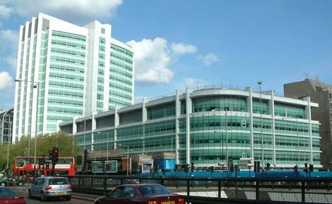
A December evening at Spitalfields Market was transformed into a hub of medical expertise during the London Pregnancy Clinic’s event. The focus was on pioneering approaches in prenatal care, particularly the 10 Week Scan,  Non-invasive prenatal testing (NIPT), and pre-eclampsia diagnosis.

Advancing the 10 Week Scan

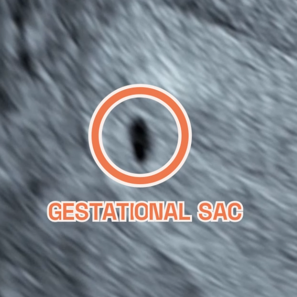
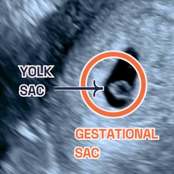
The evening began with Ms. Shaz Khojasteh, LPC’s clinical lead, greeting each attendee with a warmth that matched her expertise. Her introduction was not just an opening speech but a heartfelt welcome to LPC’s family. She shared with the audience our signature 10 Week Scan, which isn’t just a routine check. It’s a comprehensive examination of the baby’s anatomy, providing vital developmental insights at a crucial early stage, thereby enriching the information gathered before conducting NIPT.

Demystifying NIPT – Dr Fred Ushakov’s Perspective

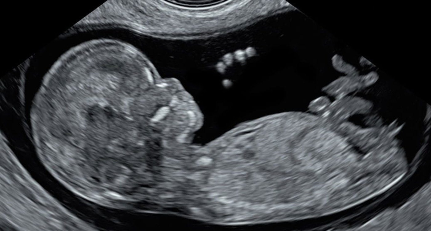
The spotlight then shifted to Dr. Fred Ushakov, whose speech was not only insightful but also visionary. He spoke passionately about his mission, in collaboration with the London School of Ultrasound and UCLH, to train the next generation of doctors and sonographers. His goal is to equip them with the skills and knowledge necessary to leverage early pregnancy scans to their full potential, enhancing patient care and pregnancy outcomes. This mission, he explained, is vital in ensuring that every pregnancy is approached with the utmost care and precision.

Dr Ushakov then addressed a common misunderstanding about NIPT. He emphasised that while NIPT is effective, it’s not all-encompassing. Patients may misinterpret it as a guarantee against all prenatal issues, not realising that conditions more severe than Down’s syndrome can occur and cannot be detected by NIPT. He further highlighted the importance of the dual approach (watch the explainer video here) that he instilled at LPC. Dr. Ushakov’s call for broader education and comprehensive scanning techniques underscored the need for a more informed approach to prenatal care.

Advocacy for Pre-eclampsia Awareness

Next, we were captivated by Dr. Daniel Stott, whose talk about pre-eclampsia was as enlightening as it was heartfelt. It was clear from his words and the energy in his voice just how dedicated he is to this topic. Dr. Stott walked us through the workings of a groundbreaking blood test for pre-eclampsia. With an approachable manner, he explained how this test isn’t just for identifying pre-eclampsia; it’s a crucial tool in carefully guiding expectant mothers through the rest of their pregnancy. 

Conclusion

As the expert talks wrapped up, the event naturally transitioned into festive networking. It was a perfect opportunity for attendees to connect, exchange experiences, and build professional ties in a holiday atmosphere. The evening emerged as a crucible of innovative ideas and shared goals for advancing prenatal care. Leaving the market, we carried with us the night’s insights and a renewed hope for the future of prenatal health. The event proved to be not just a confluence of minds but a beacon of hope for prenatal care’s future.

A special thank you to our sponsors Eurofins.

Latest Stories

Continue reading

Week 5 of Pregnancy

Graphic of Week 5 of pregnancy. An illustration of a baby in the ultrasound machine.

Week 5 of your Pregnancy

Discovering your baby’s progress since week 4

  • Published

  • Last Modified

Tags

Having journeyed through the early whispers of Week 4, we now step into the heart of your pregnancy story: Week 5. It’s a chapter filled with vibrant growth and transformation. Imagine your little one, still as tiny as an apple seed, beginning a significant journey into the embryonic period.

Week 5 of pregnancy, it’s all about growth and transformation. Your little one is busy developing, cell by cell, quickly growing from the size of a poppy seed in week 4 (watch video here) to an apple seed. We’ll explore these changes together, learn how to take care of yourself during this time and get a peek into your baby’s development with early ultrasounds. So, let’s dive into Week 5, where every day brings something new and amazing in your pregnancy journey. So let’s navigate the changes unfolding, how to care for yourself, and what to expect from medical check-ups.

Key Concepts: embryonic period

During Week 5 of your pregnancy, a crucial phase begins as your baby, the size of an apple seed, enters the embryonic period. This week marks the foundation for major organs and systems. Hormonal changes, especially increases in progesterone and human chorionic gonadotropin (hCG), are vital in supporting this stage. These hormones maintain the uterine lining and ensure the continuation of progesterone production, crucial for pregnancy health. As a result, you might notice early symptoms like fatigue and nausea. Understanding these changes is key to navigating the early stages of your pregnancy journey.

Pregnancy Checklist at 5 Weeks

While it’s still early in your pregnancy, there are some important steps you can take:

  • Confirm your pregnancy with a test if you haven’t already.
  • Schedule your first prenatal appointment to discuss your health and pregnancy. At London Pregnancy Clinic it’s called the Viability/Dating Scan. Use our pregnancy calculator to work out the best time for your scan.
  • Begin focusing on a pregnancy-safe diet and lifestyle, emphasising foods rich in folic acid and essential nutrients.

Developmental Milestones: Week 5

Growth of the Gestational Sac: This week, the gestational sac, housing your developing baby, continues to grow. It’s a clear sign of your pregnancy’s progression, providing a protective and nourishing environment for the embryo.

Yolk Sac Development: The yolk sac plays a crucial role at this stage, providing essential nutrients before the placenta is fully operational. It also contributes to the early stages of the circulatory system by producing blood cells, supporting the developing embryo’s needs. This process is essential for supporting the growing needs of your embryo and is closely tied to the growth of the gestational sac.

Heart Development: Now your baby’s heart begins its incredible journey. Initially emerging as a simple tube-like structure, it’s preparing to pulsate and evolve into a sophisticated four-chambered heart in the coming weeks. Although it’s not beating just yet, this early stage sets the stage for one of life’s most vital rhythms.

Brain and Spinal Cord Formation: Alongside the heart, the neural tube, which will become the brain and spinal cord, starts to take shape. This tube forms the bedrock of the entire nervous system, integral for everything from physical movements to cognitive functions. It’s a complex process that lays the foundation for your baby’s future mental and physical capabilities.

Emergence of Other Organs: Week 5 also marks the beginning of other organ formations. Organs like the kidneys and liver start to form, albeit in their very preliminary stages much like the heart.

What is seen on the Ultrasound: Week 5?

During the Week 5 ultrasound, expect to see the gestational sac (which has dramatically increased in size since Week 4), the first structure visible on an ultrasound, indicating your pregnancy is progressing well. This small, fluid-filled sac is where your baby will develop. Inside it, the yolk sac may also be visible, which is crucial for providing nutrients to the developing embryo and for blood cell production in these early stages. However, it’s important to remember that at this stage, the embryo itself and its heartbeat are usually not yet detectable.

It’s common not to see the embryo or hear a heartbeat during a Week 5 ultrasound. The embryo is still very small, and the heartbeat typically becomes detectable around the 6th or 7th week of pregnancy. While we know it’s tempting to have an early ultrasound, we at London Pregnancy Clinic recommend waiting until at least week 6 for your first scan. This crucial ultrasound provides a tangible glimpse into your pregnancy, offering reassurance and joy as you embark on this extraordinary journey.

  • Gestational sac at 4 weeks of pregnancy as seen on ultrasound scan.

    Gestational sac at 4 weeks of pregnancy as seen on ultrasound scan.

  • Image showing what would an ultrasound show at 5 weeks of pregnancy. It shows an Ultrasound image by London Pregnancy Clinic that shows the gestational sac and the newly developed yolk sac - for educational purpose.

    Gestational sac and yolk sac at 5 weeks of pregnancy as seen on ultrasound scan.

Thinking ahead: Week 6?

Looking forward to Week 6, expect continued development of your baby’s facial features. You may also start experiencing more pronounced pregnancy symptoms like nausea and fatigue. It’s a time to start preparing both mentally and physically for these changes.

Week 5 of Pregnancy FAQs


  • How common is it for my baby to have a heartbeat at 5 weeks?

    Your baby is an embryo (not a fetus just yet) and can have a heartbeat at 5 weeks of pregnancy, but it is not very common. The fetal heartbeat usually starts to beat between 5 and 6 weeks gestation, and it may be detectable via ultrasound around this time.

    Here are some reasons why the heartbeat might not be detected on an ultrasound at 5 weeks:

    • The embryo is too small.
    • The heartbeat is too faint.
  • What if there is no yolk sac at 5 weeks?

    Patients have asked us why is there no sac seen when 5 weeks pregnant. The yolk sac is usually visible on an ultrasound between 5 and 6 weeks gestation. If there is no yolk sac visible on an ultrasound at 5 weeks, it could be a sign of a miscarriage. 

    However, it could be a sign that the estimated gestational age is wrong. This could happen if your last menstrual period was inaccurate or if you were ovulating later than you thought.

    In this case, we may recommend a follow-up ultrasound in a week or two to see if the yolk sac has developed.


Conclusion

Week 5 of your pregnancy is a time of rapid and incredible changes. While you might start feeling different physically and emotionally, remember, these are signs of your baby’s healthy development. Stay informed, take care of yourself, and prepare for the exciting journey ahead. If you have any concerns, our team at London Pregnancy Clinic is always here to guide you.

Stay tuned on what your baby’s week-by-week growth will look like on our blog.


Latest Stories

Continue reading

Follicle Tracking Scan Explained

Graphic of Follicle Tracking Scan. An illustration of an egg in the uterus with sperm coming towards it.

Follicle tracking scan explained

Shaz Khojasteh is an advanced Specialist Sonographer in Fetal Medicine, Gynaecology and Fertility in Central London. Gynae Scans and Follicle Tracking Scans in London.

Miss Shaz Khojasteh Explains Essential Insights for Expectant Mothers.

Published

Tags

Embark on a journey of understanding with our focused insight into Follicle Tracking Scans. This blog post explores three frequently asked questions, shedding light on the significance, process, and benefits of Follicle Tracking Scans. Ideal for expectant mothers seeking early reassurance in their pregnancy, these insights aim to demystify the process and enhance your prenatal experience. Join us as we delve into the world of Follicle Tracking Scans, explained by Shaz.

Pregnancy is a time of joy and anticipation, but it also brings a plethora of questions and concerns, especially for first-time mothers. A key component of early pregnancy monitoring is the follicle tracking scan. In this blog, we explain follicle tracking scans, addressing three common questions to help expectant mothers gain clarity and confidence in their early prenatal journey.

The Basics of Follicle Tracking

Follicle tracking scans are a type of ultrasound scan used primarily in the early stages of pregnancy and fertility treatments. These scans monitor the development of ovarian follicles, which are small sacs containing eggs. Understanding follicle development is crucial for determining the best time for conception and ensuring the health of the pregnancy in its initial stages.

Importance of Follicle Tracking in Early Pregnancy

Follicle tracking is essential for those undergoing fertility treatments or trying to conceive naturally. It helps in pinpointing the ovulation period, ensuring that the timing for conception is optimal. For early pregnancy, it reassures the health and development of the embryo, offering peace of mind to expectant mothers.

Your Experience with Follicle Tracking Scans

During a follicle tracking scan, you can expect a non-invasive and painless experience. The procedure involves an ultrasound, where a small probe is used to visualise the ovaries and the developing follicles. The scan provides real-time images, allowing the healthcare professional to assess and monitor follicle growth and health. You will be asked to use our facilities and have an empty bladder for your transvaginal ultrasound scan. During your appointment, you will be provided with a skirt cover to change into. There will be a private and separate area for you to get changed. If you have specific requirements, please ask, and our team will do their best to accommodate.

Your  frequently asked questions about Follicle Tracking Scan:

What is a Follicle? A follicle is a small, fluid-filled sac located in the ovaries of the female. It plays a critical role in female fertility and the reproductive process. Each follicle contains an immature egg, or oocyte, which matures within the follicle.

Is the Follicle Tracking Scan painful? The procedure involves a qualified sonographer gently inserting a slender transvaginal ultrasound probe to obtain clear images of your ovaries. While some women might feel a slight sensation of pressure or mild discomfort, it is generally not painful.

Occasionally, mild cramping or a sense of discomfort may occur during the scan, often related to having a full bladder. This is because the bladder, when full, may exert pressure on the uterus. Rest assured, any discomfort is usually brief and tends to ease off promptly after the completion of the scan.

What affects the number of follicles? The number of ovarian follicles in women is influenced by genetics, age, medical conditions, and lifestyle factors. Genetics play a key role, as women are born with a finite number of follicles that diminish over time. Age further reduces this number, especially after the mid-30s. Medical conditions like PCOS can alter follicle count, often increasing it, which impacts ovulation and fertility. Additionally, lifestyle choices such as smoking and obesity, along with environmental toxin exposure, can negatively affect follicle numbers, potentially leading to decreased ovarian reserves and earlier menopause.

  • Graphic by London Pregnancy Clinic featuring the text 'Expert Fertility Assessment' and 'Follicle Tracking - All you need to know...' against a soft, abstract background representing fertility.

    London Pregnancy Clinic Follicle Tracking Overview

    London Pregnancy Clinic Follicle Tracking Overview

  • Informative illustration by London Pregnancy Clinic explaining follicle tracking scans with labeled diagram of the ovary showcasing different stages from the follicle phase to the luteal phase.

    What is Follicle Tracking?

    What is Follicle Tracking?

  • Detailed explanation by London Pregnancy Clinic of what sonographers look for during a follicle tracking ultrasound scan, including the number, size, and maturity of follicles.

    Follicle Tracking Details

    Follicle Tracking Details

  • Educational content by London Pregnancy Clinic indicating the optimal timing for follicle tracking scans during the menstrual cycle and for IVF treatments, with a circular diagram of the menstrual cycle phases.

    Best Time for Follicle Tracking

    Best Time for Follicle Tracking

  • Graphic by London Pregnancy Clinic outlining the reasons for follicle tracking, noting its non-invasive nature and its role in identifying the best times for fertility treatments and natural conception.

    Purpose of Follicle Tracking

    Purpose of Follicle Tracking

  • Next steps guide by London Pregnancy Clinic after a follicle tracking scan, advising patients on discussing results with their clinicians and offering additional consultation services for a fee.

    Next Steps After Follicle Tracking

    Next Steps After Follicle Tracking

  • Ms. Shahrzad Khojasteh, Clinical Lead and Specialist Sonographer at London Pregnancy Clinic, smiling in professional attire with text highlighting services in fertility screening, including follicle tracking, endometrial lining, and pregnancy scans.

    Specialist Sonographer

    Specialist Sonographer

Conclusion

Understanding the role and process of Follicle Tracking Scans is a vital part of your pregnancy journey. These scans offer invaluable insights into the early stages of pregnancy, aiding in conception and providing reassurance. If you have more questions or need personalised advice, we invite you to contact the London Pregnancy Clinic for more information.

Latest Stories

Continue reading

Week 4 of Pregnancy

Graphic of Week 4 of pregnancy. An illustration of a zygote in the uterus.

Week 4 of your Pregnancy

Your Baby’s Remarkable Beginning

  • Published

  • Last Modified

Tags

This blog post is part of a series that breaks down early pregnancy week by week. Today we will be talking about Week 4 of your pregnancy journey! Within this article, we’ll delve into the astonishing advancements occurring during this initial phase of pregnancy. Although your baby remains incredibly minuscule, measuring at no more than 1 millimeter (similar to a poppy seed), a realm of growth and transformation is already well underway.

Key Concepts: Gestational Weeks vs. Post-Conception Weeks

Understanding the difference between gestational weeks and post-conception weeks is crucial for accurately tracking the progress of your pregnancy.

Gestational Weeks: Gestational age refers to the age of the pregnancy and is measured from the first day of the mother’s last menstrual period (LMP). This is the standard method used by healthcare providers to track pregnancy. For example, at 4 weeks gestation, the embryo is about 2 weeks post-conception. Gestational age is crucial for determining the due date and monitoring the baby’s development.

Post-Conception Weeks: Post-conception age, also known as embryonic age, is measured from the time of conception. It provides a more precise timeline of the baby’s development. For instance, at 4 weeks post-conception, the embryo is undergoing significant developments, such as the formation of the placenta and the beginning of the amniotic sac.

Pregnancy Checklist at 4 Weeks

While it’s still early in your pregnancy, there are some important steps you can take:

  • During the initial 4 weeks of pregnancy, you may not experience noticeable symptoms. To verify your pregnancy, consider taking a pregnancy test.
  • Once you’ve confirmed your pregnancy you may think about your first prenatal visit with your doctor, even though it may not occur for another couple of weeks. With us, this would be our Viability/Dating Scan. During this visit, our specialists, like Miss Shaz Khojasteh, will check for single/multiple pregnancies, date the pregnancy and check the main structures of the gestational sac. We believe early pregnancy scans are essential for peace of mind and early reassurance for parents. 
  • Focus on adopting healthy diet and lifestyle habits. Say goodbye to smoking and alcohol, opt for nutritious foods, and stay well-hydrated.
  • The NHS provides valuable recommendations for expectant mothers, including considering the importance of taking supplements during pregnancy. Additionally, it’s essential to recognise how emotional changes that often accompany pregnancy can impact your relationships.

Developmental Milestones: Week 4

By Week 4, your baby develops from being a single-cell zygote to a blastocyst. Rapid cell division shapes the future. At this stage, your embryo completes its journey from the fallopian tube to the uterus, burrowing into the uterine lining. Half becomes your future child, and the other forms the placenta—a vital nutrient carrier.

The amniotic sac, often called the “bag of waters,” forms around the embryo, along with the yolk sac, important for your baby’s digestive system.

Your embryo now consists of three unique cell layers:

  • The endoderm—nurturing your baby’s digestive system, liver, and lungs.
  • The mesoderm—laying the foundation for your baby’s heart, sex organs, bones, kidneys, and muscles.
  • The ectoderm—shaping your baby’s nervous system, hair, eyes, and outer skin layer.

What is seen on the Ultrasound: Week 4?

One of the common queries we receive at the London Pregnancy Clinic is whether having an ultrasound at 4 weeks gestation is a requirement. At this stage, around the fourth week of pregnancy, significant developments are occurring. The blastocyst is in the process of dividing into an embryo and placenta. However, it’s crucial to understand that an ultrasound of your uterus during this early stage will typically reveal what appears to be a minuscule dot known as the gestational sac, and it’s important to note that a heartbeat is not typically detectable at this early point of development. Detecting a more advanced pregnancy may require waiting until a later stage.

Thinking ahead: Week 5?

The next steps in your pregnancy journey involve preparing for your first ultrasound appointment. It’s completely normal to eagerly anticipate your first ultrasound, but it’s essential to be aware that at 4 and 5 weeks of gestation, the gestational sac is typically too small to be easily visible on the scan. For a clearer image of your pregnancy, it is generally advisable to wait until around 6 weeks or later, when the development progresses. However, it’s worth noting that in certain cases, skilled sonographers with extensive experience may be able to discern subtle indications of pregnancy by closely observing changes in the uterine lining. You can use our Pregnancy Calculator to work out your optimal scan date range by entering your Last Menstrual Period(LMP).

Week 4 of Pregnancy FAQS?


  • Is 4 weeks pregnant too early to test

    At 4 weeks, it’s not too early to take a pregnancy test. Most home pregnancy tests are designed to be sensitive enough to detect the pregnancy hormone, human chorionic gonadotropin (hCG), in your urine by this time. This is typically around the time your period is due, which is when hCG levels are sufficiently high to indicate pregnancy.

    While you can confirm pregnancy with a test and even start calculating your due date, it’s indeed too early for an ultrasound scan. At 4 weeks, the baby is very small, and significant developmental milestones like the heartbeat, which typically becomes detectable around 6 weeks of gestation, have not yet occurred. Therefore, while a home pregnancy test can provide early confirmation, an ultrasound scan at this stage wouldn’t offer much information. For more detailed and visual insights into your baby’s development, waiting until at least the 6th week or later is advisable when the heartbeat and other embryonic structures start to become visible.

  • What to do when you’re 4 weeks pregnant?

    • Confirm Your Pregnancy: A home pregnancy test can confirm your suspicion of being pregnant. It’s best to take the test after you’ve missed your period for the most accurate result.

    • Schedule a Doctor’s Appointment: Even though it’s early, getting in touch with a healthcare provider is a good idea. They can confirm your pregnancy through a blood test and start guiding you through prenatal care.

    • Document Your Journey: Consider starting a pregnancy journal or diary to record your experiences, thoughts, and feelings. This can be a wonderful keepsake for the future.

    • Rest and Relax: Early pregnancy can bring fatigue and other symptoms. Ensure you’re getting enough rest and practising stress-relief techniques.

  • What does 4 weeks pregnancy look like?

    At 4 weeks of pregnancy, external changes to your body are typically minimal and not yet noticeable to others. However, internally, a remarkable series of events is unfolding. Your embryo, though only about the size of a poppy seed, is rapidly developing. This tiny cluster of cells is already beginning the incredible process of forming what will become vital organs and structures.


Conclusion

Week 4 of your pregnancy signifies the remarkable commencement of your baby’s journey. While ultrasound visuals may not reveal much at this early stage, rest assured that a realm of development is quietly unfolding within your body. Join us as we look forward to exploring the upcoming stages of your pregnancy journey, complete with the changes and milestones that await.

It’s crucial to remember that every pregnancy is unique. Therefore, we recommend consulting your healthcare provider for tailored guidance and attentive care during this special period. Our team is here to provide support throughout your exciting pregnancy journey!

Once again, heartfelt congratulations on your pregnancy. Stay tuned for further updates as we continue to monitor your baby’s week-by-week growth.


Latest Stories

Continue reading

UCLH: A Beacon of Excellence in Obstetrics and Gynaecology

UCLH: A Beacon of Excellence in Obstetrics and Gynaecology

Published

Tags

University College London Hospitals NHS Foundation Trust (UCLH) stands as a paragon of excellence in the healthcare sector, particularly in obstetrics and gynaecology. With a legacy rooted in innovation and top-tier medical services, UCLH has garnered global recognition, ranking among the world’s best in its field. Beyond its renowned maternity services and the esteemed Fetal Medicine Unit (FMU), UCLH’s partnership with the London Pregnancy Clinic showcases a team of distinguished professionals dedicated to women’s health. Together, they underscore UCLH’s unwavering commitment to providing unparalleled care for women at every life stage.

University College London Hospitals NHS Foundation Trust (UCLH) has long been recognized as a leading institution in the realm of healthcare. With a rich history and a commitment to innovation and excellence, UCLH has consistently delivered top-tier medical services to its patients. One area where UCLH has particularly excelled is in the field of obstetrics and gynaecology.

Global Recognition

According to the Newsweek’s World’s Best Specialized Hospitals 2024 rankings, UCLH is among the top hospitals globally for obstetrics and gynaecology. This recognition is a testament to the hospital’s dedication to providing the best care for women at every stage of their lives.

Gynaecology at UCLH

The gynaecology service at UCLH offers a comprehensive range of treatments and services. From general gynaecological care to specialized treatments for conditions like endometriosis and fibroids, the department is equipped to handle a wide array of women’s health issues. The team also provides support for early pregnancy and acute gynaecological problems.

Maternity Services

UCLH’s maternity services are designed to support women throughout their pregnancy journey. The hospital offers everything from antenatal classes to postnatal support, ensuring that mothers and their babies receive the best care possible.

A standout feature of UCLH’s maternity services is the Fetal Medicine Unit (FMU). Led by a team of highly skilled specialists, the FMU offers diagnosis and treatment for complications that may arise in unborn babies. Recognized both nationally and internationally, the FMU sees over 7,000 patients each year, providing care in a supportive and professional environment.

A Rich History and a Bright Future

The University College London Hospitals NHS Foundation Trust has a storied history that dates back to the 18th century. Over the years, the trust has grown and evolved, incorporating various hospitals and expanding its range of services. Today, UCLH is not just a hospital but a conglomerate of medical institutions dedicated to research, teaching, and patient care.

In partnership with University College London, UCLH has major research activities, further cementing its reputation as a leading institution in the medical field. Each year, its hospitals treat over 500,000 outpatients and admit over 100,000 patients, showcasing the trust’s vast reach and impact.

The London Pregnancy Clinic Connection

At London Pregnancy Clinic, we are proud that many of our practitioners and support staff are either currently practicing at UCLH or have been trained there:

Dr. Fred Ushakov

Dr. Fred Ushakov is the Founder and Medical Director of the London Pregnancy Clinic. With over 35 years of medical experience, he specializes in fetal medicine and ultrasound. Notably, he is recognized as one of the most skilled ultrasound operators at the UCLH Fetal Medicine Unit. Dr. Ushakov’s contributions extend beyond clinical practice; he founded the London School of Ultrasound and the Early Fetal Scan Conference. His dedication to the field is evident in his role as an Ambassador of the International Society of Ultrasound in Obstetrics and Gynaecology (ISUOG) from 2014-2020.

Ms Shahrzad (Shaz) Khojasteh

Ms Shaz Khojasteh is a Specialist Sonographer in Obstetrics and Gynaecology. She completed her Master’s degree in Prenatal Genetics and Fetal Medicine at UCL in 2016 and underwent specialized training in Fetal Medicine Ultrasound at Kings College Hospital. Shaz has also received training in Fetal Echocardiography and has gained valuable experience in Early Pregnancy and Gynaecological ultrasound scanning.

Dr. Giovanni Granozio

Dr. Giovanni Granozio is a clinical fellow in fetal medicine at the Royal London Hospital and consults privately at the London Pregnancy Clinic. He specializes in pregnancy screening scans, particularly for pregnancies with fetal anomalies, growth-restricted babies, and twin pregnancies. Dr. Granozio graduated from the University of Salerno and later specialized in Gynaecology and Obstetrics at the University of Turin. He relocated to London in 2020 to further his training in Fetal Medicine and Obstetric Ultrasound at UCLH.

Dr. Daniel Stott

Mt Stott is a distinguished consultant in obstetrics, practicing at The Portland Hospital and University College Hospital London (UCLH). He has notably established a successful antenatal clinic for women with hypertension at UCLH and serves as the Trust’s clinical lead for foetal monitoring. Dr. Stott’s expertise lies in maternal and fetal medicine, with a particular focus on obstetric ultrasound scanning, prenatal diagnosis, and maternal medicine. He has a keen interest in pregnancies complicated by hypertension and in fetal medicine. His academic pursuits are evident in his role as an honorary lecturer at UCL and his extensive research publications in obstetric and gynaecology journals.

Latest Stories

Continue reading

The History of Ultrasound in Obstetrics and Gynaecology

The History of Ultrasound in Obstetrics and Gynaecology

Published

Tags

The world of medical imaging has seen transformative technologies over the years, and ultrasound stands tall as one of the most pioneering. This non-invasive imaging tool has become an indispensable asset in fetal medicine and gynaecology. Let’s delve deep into the history of ultrasound, understanding its workings and establishing its safety credentials.

The Dawn of Ultrasound in Medicine

The journey of ultrasound began in the early 20th century. Initially, it was utilised for industrial and marine purposes, primarily to detect submarines. It wasn’t until the 1950s that scientists began to recognise its potential in medical diagnostics. The foundational use in obstetrics was to detect and measure foetal size, growth and position, making it a pivotal tool for doctors.

Ultrasound’s Ascent in Fetal Medicine

The 1970s and 1980s were transformative decades for ultrasound. As technology advanced, the clarity and details of the ultrasound images improved exponentially. This led to its expanded role in foetal medicine. Doctors could now not only measure the size and position of the foetus but also identify structural abnormalities, understand foetal behaviour, and estimate gestational age with enhanced precision.

Screening for Down’s syndrome, congenital heart diseases, and neural tube defects became possible, marking ultrasound’s vital role in antenatal care. The ability to visualise the foetus in the womb has not only improved clinical outcomes but also allowed parents to establish an early bond with their unborn child.

Ultrasound’s Role in Gynaecology

Ultrasound in gynaecology has been revolutionary. From visualising ovarian cysts to diagnosing endometriosis, it offers a pain-free, non-invasive solution for women. The introduction of transvaginal ultrasound probes in the 1980s provided clearer, more detailed images of the uterus and ovaries, enhancing diagnostic precision.

How Does Ultrasound Work?

In layman’s terms, ultrasound uses high-frequency sound waves to produce images of structures inside the body. A device called a transducer is placed on the body, and it emits sound waves. When these waves hit a boundary between tissues, like between fluid and soft tissue, they bounce back. The returning echoes are translated by a computer into images displayed on a screen.

Is Ultrasound Safe for Humans?

One of the paramount reasons for ultrasound’s popularity is its safety. Unlike X-rays, ultrasound doesn’t use radiation. Over decades of use, there’s been no concrete evidence linking ultrasound to any harmful side effects, making it a preferred choice for examining pregnant women and their unborn babies.

However, like any medical procedure, it should be used judiciously and only when medically necessary. It’s comforting for patients to know that they’re in safe hands when undergoing an ultrasound.

In Conclusion
From its marine roots to the pinnacle of medical diagnostics, ultrasound has traversed a fascinating journey. Today, it stands as an emblem of innovation in fetal medicine and gynaecology, providing invaluable insights while ensuring patient safety. As technology continues to evolve, the horizon for ultrasound promises even more groundbreaking discoveries.

Latest Stories

Continue reading

A Comprehensive Guide to AMH Blood Test: Its Significance and Connection to IVF

Ever wondered how a simple blood test could offer a glimpse into your fertility? In this blog post, we’ll delve into one of the most crucial hormones affecting fertility—the Anti-Mullerian Hormone (AMH). If you’ve heard of this hormone and wondered what role it plays in fertility or IVF treatments, you’re in the right place.

What is the AMH Blood Test?

An AMH (Anti-Mullerian Hormone) blood test measures the levels of AMH in a woman’s blood. Produced by the cells of the ovarian follicles, AMH serves as a reliable marker of ovarian reserve, effectively indicating a woman’s fertility potential. This test aids clinicians and fertility specialists in predicting a woman’s response to fertility treatments, particularly in vitro fertilisation (IVF).

What is the Anti-Mullerian Hormone (AMH)?

The Anti-Mullerian Hormone, first discovered by Alfred Jost in the 1940s, plays a crucial role in the sexual development of foetuses. AMH was initially known for its role in male sexual development, but subsequent research has unearthed its pivotal part in female fertility. In women, the hormone is released by the antral follicles, the small sacs in the ovaries that hold the eggs.

Why is the AMH Blood Test Important?

As AMH levels correspond to the number of remaining eggs in the ovaries, the AMH blood test offers a reliable gauge of a woman’s ovarian reserve. However, it’s essential to understand that AMH levels do not directly correlate with the quality of eggs.

Low AMH levels might indicate a diminished ovarian reserve, which could be a concern for women attempting to conceive naturally or through IVF. In contrast, higher AMH levels may suggest conditions like Polycystic Ovary Syndrome (PCOS).

When is an AMH Blood Test Required?

The AMH test may be recommended in several circumstances:

  1. Evaluating Ovarian Reserve: AMH levels help assess the number of eggs remaining in the ovaries. The test may be suggested for women approaching their mid-30s, those with a family history of early menopause, or those who’ve previously undergone chemotherapy or pelvic surgery.
  2. Diagnosing PCOS: Elevated AMH levels may be indicative of PCOS, a common condition affecting a woman’s ovaries and ovulation.
  3. Determining IVF Success: Before starting IVF treatment, clinicians may order an AMH test to better predict how the ovaries will respond to the fertility drugs used to stimulate egg production.

Connection between AMH Levels and IVF

The AMH blood test holds significant value in the field of assisted reproductive technology, especially IVF. It helps clinicians devise personalised treatment strategies for their patients.

For women with low AMH levels, special considerations may be needed during the stimulation phase of IVF. These women often respond less to ovarian stimulation, leading to a smaller number of eggs retrieved, which can affect the success rate of IVF. Conversely, women with high AMH levels might be at risk for Ovarian Hyperstimulation Syndrome (OHSS), a condition that can occur when the ovaries are overly stimulated by fertility drugs.

When is the Best Time to Have an AMH Blood Test?

An AMH blood test can be performed at any time during a woman’s menstrual cycle, as AMH levels remain relatively constant throughout. However, many fertility clinics might suggest the test as a part of an initial fertility assessment or prior to beginning fertility treatments like IVF.

In Conclusion

The AMH blood test serves as a potent tool in assessing a woman’s ovarian reserve and planning personalised fertility treatments. Whether you’re attempting to conceive naturally, preparing for IVF, or just wish to know more about your fertility, an AMH test could offer invaluable insights. Always consult with a fertility specialist to discuss your options and understand what’s best for your individual circumstances.

Discover more about the Blood Tests we offer at London Pregnancy Clinic. Book your AMH Test today for £80.

Pregnancy Testing – Deep Dive into the Beta HCG Test

Pregnancy tests aim to provide a simple yet effective method of determining if you’re pregnant. There are two primary types of pregnancy tests available: urine tests and blood tests. While urine tests are more commonly used due to their convenience and over-the-counter availability, blood tests, specifically the Beta HCG (Human Chorionic Gonadotropin) test, offer a more detailed insight into the pregnancy. Here’s an in-depth look at the Beta HCG pregnancy test, how it differs from urine tests, its accuracy, uses, and how to interpret the results.

What is a Beta HCG Pregnancy Test?

A Beta HCG test is a blood test used to detect pregnancy. Human Chorionic Gonadotropin (HCG) is a hormone produced by the placenta after implantation. The beta subunit of HCG can be detected in the blood serum of expectant mothers approximately 11 days after conception; HCG can be detected in the urine 12-14 days after conception.

There are two types of HCG tests:

  1. Qualitative HCG test: This test simply detects the presence of HCG in the blood. It gives a “yes” or “no” answer to the question, “Are you pregnant?” Doctors often order this test to confirm pregnancy.
  2. Quantitative HCG test (Beta HCG): This test measures the specific level of HCG in the blood. It can detect even very low levels of HCG, making it a highly accurate test.

How Does a Beta HCG Test Differ from Urine Tests?

While both urine pregnancy tests and Beta HCG tests aim to detect the presence of HCG, there are distinct differences:

  1. Method of Testing: Urine tests can be done at home, while Beta HCG tests require a blood sample taken in a healthcare setting.
  2. Sensitivity: Beta HCG tests are more sensitive than urine tests and can detect lower levels of HCG. This means they can confirm pregnancy earlier than urine tests — typically around 11 days after conception.
  3. Information Provided: While urine tests only provide a positive or negative result, a Beta HCG test can provide more information about the pregnancy, like the approximate age of the fetus, or if there are potential complications.

The Accuracy of Beta HCG Pregnancy Test

Beta HCG tests are highly accurate. Because they can measure the exact amount of HCG in the blood, they can detect pregnancy earlier than urine tests, and they can also confirm or rule out potential problems.

However, while the Beta HCG test can detect if you’re pregnant, a single test can’t always accurately predict a healthy pregnancy. Levels of HCG can vary greatly among individuals, and a single measurement isn’t always indicative of a problem or complication. Doctors often will test HCG levels more than once, looking at how the levels change over time.

Uses of Beta HCG Pregnancy Test

Aside from confirming pregnancy, a Beta HCG test can be used for the following:

  1. Monitoring Pregnancy: Doctors may use Beta HCG tests to monitor the health of a pregnancy, particularly in the early weeks. They may use serial measurements to see if HCG levels are rising as they should.
  2. Identifying Ectopic Pregnancy: Lower than usual levels of HCG or levels that do not increase as they should may suggest an ectopic pregnancy — where the fertilized egg implants outside the uterus.
  3. Screening for Down’s Syndrome: As part of the “double,” “triple,” or “quadruple” screen test, the Beta HCG test can help assess a baby’s risk of Down syndrome.

Interpreting Beta HCG Test Results

Interpreting Beta HCG test results isn’t always straightforward, as normal HCG levels can vary widely. Typically, in a healthy pregnancy, HCG levels double approximately every 48 hours in the first weeks following conception.

However, unusually high or low levels, or levels that don’t increase as they should, might indicate a problem, such as an ectopic pregnancy, a miscarriage, or a potential chromosomal abnormality. In such cases, your healthcare provider will likely order further tests to determine the cause of the abnormal HCG levels.

It’s important to remember that while the Beta HCG test is a crucial tool in pregnancy monitoring, it is just one aspect of a larger picture. It should be used in conjunction with other tests and examinations to ensure a comprehensive understanding of both maternal and fetal health.

In conclusion, the Beta HCG pregnancy test is a sensitive, accurate, and versatile tool in the early detection and monitoring of pregnancy. By understanding its uses and how to interpret the results, you can gain a more comprehensive view of your pregnancy journey.

Here are typical ranges for hCG levels during pregnancy:

  • 3 weeks: 5 – 50 mIU/mL
  • 4 weeks: 5 – 426 mIU/mL
  • 5 weeks: 18 – 7,340 mIU/mL
  • 6 weeks: 1,080 – 56,500 mIU/mL
  • 7 – 8 weeks: 7,650 – 229,000 mIU/mL
  • 9 – 12 weeks: 25,700 – 288,000 mIU/mL
  • 13 – 16 weeks: 13,300 – 254,000 mIU/mL
  • 17 – 24 weeks: 4,060 – 165,400 mIU/mL
  • 25 – 40 weeks: 3,640 – 117,000 mIU/mL

Looking to book a blood test with us? Please visit our Blood Tests page. If you already had your pregnancy confirmed, we recommend to book an early pregnancy scan to check for the baby’s wellbeing.

Your Fertility Scan Guide 

Endometrial Lining Scan 

Endometrial Lining Scan, also known as the ‘Baseline Scan’, is designed to assess the thickness of the womb as well as its general outline and structure. 

These scans are popular amongst patients going through IVF treatment and are designed to guide your referring clinician, helping them to understand and plan your treatment accordingly. This is done by estimating the best time and chances for conception in addition to preparation prior to embryo transfer. Book here.

London Pregnancy Clinic offer three price packages for Endometrial Lining Scans: 

  • Endometrial Lining Scan/Baseline Scan – £120 
  • One lining ultrasound scan 
  • Rose Package – £220 
  • Two lining ultrasound scans 
  • Lily Package – £320 
  • Three lining ultrasound scans 

Follicular Tracking Scan 

Follicular Tracking Scan, also known as Antral Follicle Count, assesses the total number and size of the follicles in each ovary. 

These scans are popular amongst those going through IVF treatment. Follicle Tracking Scan helps your referring clinician to make important decisions in your fertility treatment and as such it is imperative to get accurate readings. Book now.

Similar to Endometrial Lining Scan, we offer three price packages for Follicular Tracking Scans: 

  • Follicle Tracking Scan/Antral Follicle Count: £150 
  • One follicle tracking ultrasound scan 
  • Rose Package – £270 
  • Two follicle tracking ultrasound scans 
  • Lily Package – £390 
  • Three follicle tracking ultrasound scans 

HyCoSy Scan 

This is an advanced ultrasound scan to examine the fallopian tubes, uterus and ovaries providing an important insight into your fertility. 

HyCosy scans are performed by our expert Consultant Gynaecologist, Mr Prashant Purohit, specialises in reproductive medicine and fertility assessment. HyCoSy scans are recommended for patients who have experienced difficulties in getting pregnant. Book now.

  • Cost: £500 including a pre-procedure consultation 

Blood Test Services 

London Pregnancy Clinic offers a variety of different blood tests with a short turnaround time. The results will be sent to you and your referring consultant through our secure online system. 

  • 1
  • 2