What Is Cryptic Pregnancy?

What Is Cryptic Pregnancy?
Understanding the Phenomenon of Silent Pregnancy
-
Published
-
Last Modified
Tags
Cryptic pregnancy is a fascinating phenomenon that has puzzled both medical professionals and expecting mothers alike. In this blog post, we will delve into hidden pregnancy, exploring its definition, signs and symptoms, causes, emotional impact, and more. Whether you’re an expecting mother or simply curious about pregnancy-related topics, join us on this enlightening journey.
It’s estimated that approximately 1 in 475 women will experience a cryptic pregnancy until 20 weeks of gestation. Hidden pregnancy is a condition where a woman remains unaware of her pregnancy until very late in the gestation period or even until delivery. While it sounds like something out of a movie, cryptic pregnancy is a real and relatively rare phenomenon. Despite its rarity, the impact on those who go through it can be significant.
What is Cryptic Pregnancy?
Cryptic pregnancy, also known as stealth pregnancy or denial of pregnancy, refers to a pregnancy that goes undetected by the woman herself and often by healthcare professionals as well. The reasons behind this lack of awareness vary, but it’s important to understand that it’s not a conscious decision to deny or ignore the pregnancy. It is characterised by irregular or absent menstrual periods, minimal or no pregnancy symptoms, and negative pregnancy tests.
Signs and Symptoms of Cryptic Pregnancy
One of the most intriguing aspects of cryptic pregnancy is the absence of typical pregnancy symptoms. While some women may experience certain symptoms such as weight gain, abdominal distension, or intermittent bleeding, many others will have no noticeable signs of pregnancy. The lack of these symptoms, combined with irregular menstrual cycles, can contribute to the confusion and disbelief surrounding this type of pregnancy.
Indicators to look out for
- Missed period: While most individuals recognise pregnancy due to a missed period, this is not always the case. If your periods are irregular, you may not notice a missed period as a sign of pregnancy. However, people may experience spotting or light bleeding, which can be mistaken for a period.
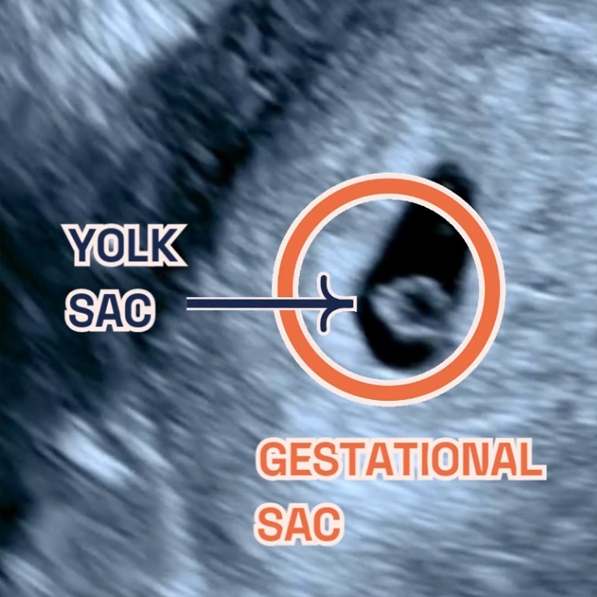
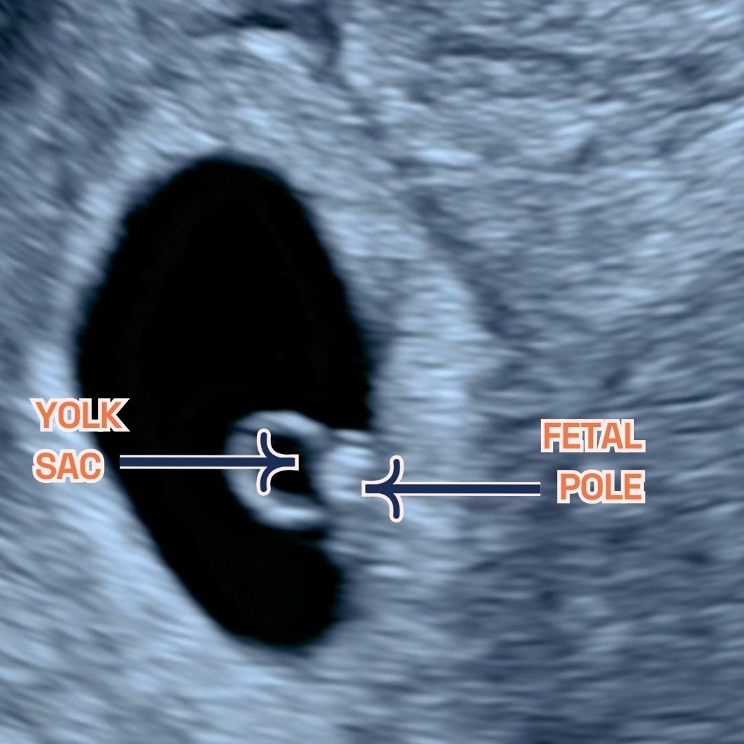
- Fetal movement: Most individuals do not feel the fetus move until about halfway through pregnancy, so around the middle of the second trimester. If you have an anterior placenta (located at the front of your belly), it may be more challenging to perceive kicks. In the case of cryptic pregnancy, this could be why you are unaware of being pregnant.
- False negative pregnancy test result: This occurs when someone is actually pregnant, but the test indicates otherwise. If you do not use an at-home pregnancy test correctly, you may receive an inaccurate result. To be certain, it is always best to consult a healthcare provider to confirm pregnancy. Another way you can check whether you’re pregnant or not is by doing one of the pregnancy blood tests.
- Weight gain or swollen belly: Some individuals may not realise they have gained weight due to pregnancy, or they may attribute an unusually bloated belly to a particular food.
Please note that this response does not constitute medical advice. If you have concerns or questions about pregnancy, consult a qualified healthcare professional.
Factors Contributing to Cryptic Pregnancy
Various factors, both physiological and psychological, can influence cryptic pregnancy. Hormonal imbalances or irregularities can disrupt the usual hormonal markers of pregnancy and lead to false-negative pregnancy tests. Certain medical conditions, such as polycystic ovary syndrome (PCOS) or uterine abnormalities, can also affect the accuracy of pregnancy tests. Additionally, psychological factors, such as subconscious denial or a lack of awareness about pregnancy symptoms, can contribute to a woman’s unknowingness.
Potential complications of a cryptic pregnancy?
Complications arising from a cryptic pregnancy stem from the lack of awareness about one’s condition. These complications may include:
- Absence of prenatal care: Unaware of the pregnancy, individuals do not receive the necessary prenatal care, such as vital bloodwork, nutritional guidance, ultrasounds, and other essential support required for a healthy pregnancy. Furthermore, not knowing about the pregnancy could result in unassisted labour without the aid of skilled healthcare professionals.
- Necessity for lifestyle adjustments: Smoking and consuming alcohol have detrimental effects on pregnancy. In the case of a cryptic pregnancy, individuals may unknowingly engage in these harmful practices or use medications and supplements that are generally unsafe for expectant mothers.
- Elevated risk of medical conditions: Without proper care or diagnostic tests, conditions like gestational diabetes or preeclampsia may go undetected.
- Heightened risk of congenital conditions: The fetus is at a higher risk of developing congenital conditions due to the lack of genetic tests, like NIPT, or other evaluations that aid in diagnosing the health of the baby.
It is crucial to highlight the potential risks associated with a hidden pregnancy to promote awareness and encourage individuals to seek appropriate medical support. This fact has been highlighted in studies saying that infants born from cryptic pregnancies are more likely to be born prematurely, placing them at risk of poor growth and respiratory issues.
Emotional Journey of Cryptic Pregnancy
The emotional journey of hidden pregnancy is complex and can vary greatly from woman to woman. Discovering that you are pregnant after an extended period of unknowingness can evoke a range of emotions, including shock, confusion, and even joy. It’s crucial for women experiencing cryptic pregnancy to seek emotional support from their partners, family, and healthcare professionals. Counselling and therapy can also be beneficial in navigating the emotional challenges that may arise.
Cryptic Pregnancy FAQs
-
Can stealth pregnancy be prevented?
Cryptic pregnancy, also known as hidden pregnancy, cannot be prevented as it is often caused by physiological or psychological factors that are beyond an individual’s control.
-
Can cryptic pregnancy occur in women who have regular periods?
Yes, cryptic pregnancy can occur in women with irregular or regular menstrual cycles. Irregular periods do not necessarily indicate cryptic pregnancy, and regular periods do not guarantee the absence of pregnancy.
It’s important to consult with a healthcare professional, such as a gynaecologist, for accurate information and personalised insights into cryptic pregnancy.
Expert Opinion: For professional insights into the medical aspects of cryptic pregnancy, including causes, symptoms, and diagnosis, you can talk to our in-house gynaecologist Dr. Prashant Purohit. Alongside LPC, he is a Consultant in Obstetrics, Gynaecology, and Reproductive Medicine based at Kings College Hospital NHS Foundation Trust and Kings Fertility, London. He specialises in the management of infertile couples, fertility preservation, fibroids, endometriosis, miscarriage, polycystic ovaries, menopause, menstrual bleeding disorders, and pelvic pain. His special interests include minimally invasive surgery like (key-hole) laparoscopic/ hysteroscopic surgery, Gynecological and early pregnancy ultrasound, and reproductive medicine.
Conclusion
Cryptic pregnancy is a unique and captivating phenomenon that challenges our understanding of pregnancy and the human body. While it may be rare, it’s important to acknowledge and support individuals who experience cryptic pregnancy, as their journey can be filled with confusion, uncertainty, and emotional complexity. By raising awareness and fostering understanding, we can create a more inclusive and supportive environment for all women navigating the intricacies of pregnancy.