
Author: London Pregnancy Clinic

NIPT and Scan: Why We Champion This Dual Approach

Graphic of a baby footprint in a heart with DNA strand on either side.

NIPT and Scan Approach: Why We Champion This Screening Method at London Pregnancy Clinic

Published

Tags

At the London Pregnancy Clinic, we’re dedicated to providing expectant mothers with advanced screening options and the highest level of care. We firmly recommend combining Non-Invasive Prenatal Testing (NIPT) and Ultrasound screening. Let’s explore why we endorse NIPT and Scan approach and how it benefits our patients.

Why Choose Both NIPT and scan?

Comprehensive Screening:  

Ultrasound visually assesses the baby’s anatomy, checking for physical abnormalities and measuring growth. NIPT, known by brand names like Natera’s Panorama AI or Eurofins’ PrenatalSafe, examines fetal DNA in the mother’s bloodstream, providing insights into potential chromosomal abnormalities like Down’s Syndrome, Edwards syndrome, and Patau syndrome.

Increased Accuracy and Early Detection:  

By merging Ultrasound’s structural insights with genetic data from NIPT, we significantly reduce false positives and offer more accurate results. As early as 10 weeks, when your baby is the size of a strawberry, we initiate the dual screening process. At this stage, we conduct the earliest possible structural anomaly scan, the Ten-week Anomaly Scan, to search for structural anomalies that NIPT can’t detect. We can rule out severe physical abnormalities like Acrania, Spina bifida, Absence of arms, hands, legs or feet, and Alobar holoprosencephaly. Only after confirming your baby’s structural development do we proceed with the NIPT test.

UNDERSTANDING THE TECHNOLOGY

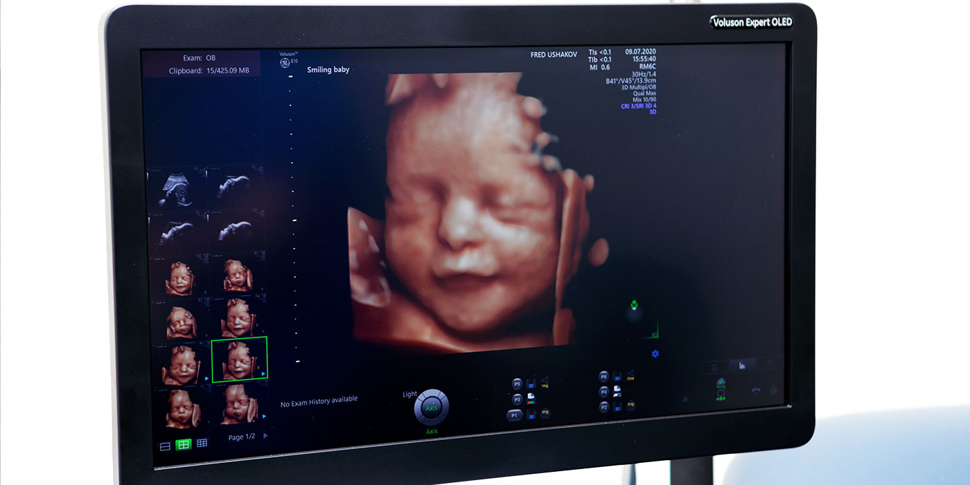
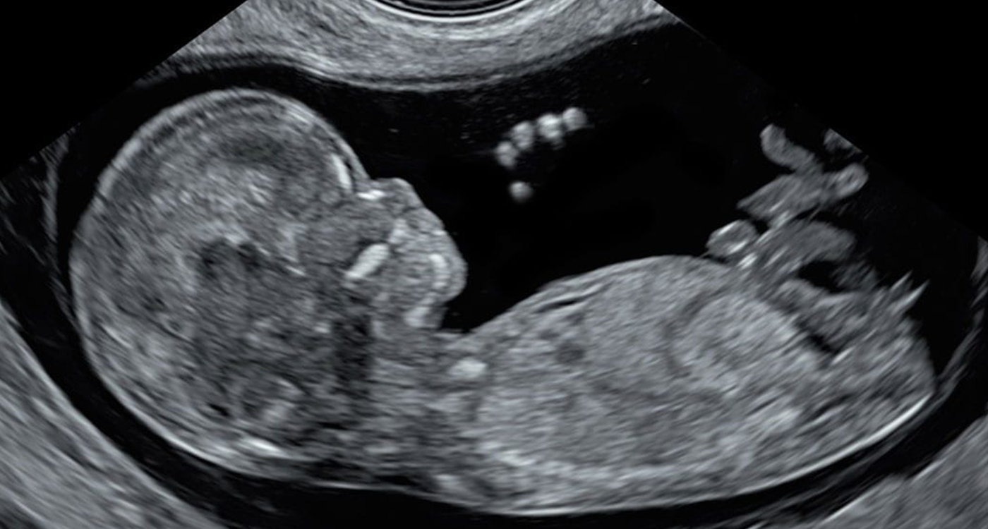
Ultrasound Screening: 

Ultrasound employs sound waves to create images of the baby in the womb. A small probe, called a transducer, moves over the mother’s abdomen. The transducer emits high-frequency sound waves that bounce off the baby’s structures, and these echoes are converted into images on a screen.

Non-Invasive Prenatal Testing (NIPT): 

NIPT is a simple blood test taken from the expectant mother. This test detects tiny fragments of the baby’s DNA circulating in the mother’s bloodstream. By analysing these fragments, we can determine the risk of certain chromosomal conditions.

Is It Safe?

Absolutely. Both Ultrasound and NIPT are non-invasive and pose minimal to no risk to both mother and baby. However, it’s important to note that while NIPT is highly effective, it’s not a definitive diagnostic test. In cases of low negative predictive value, our doctors may recommend invasive tests like CVS or amniocentesis, which carry minimal miscarriage risk.

Our NIPT Options

As early as…
  • 10 weeks

  • 9 weeks

  • 10 weeks

Turnaround (Working Days)
  • 2-4

  • 5-7

  • 5-7

Lab Location
  • UK

  • US

  • US

No Call Results
  • <1%

  • <1%

  • <1%

Redraw Rate
  • 2%

  • 3%

  • 2%

Edward’, Patau & Down’s Syndrome
  • ✔

  • ✔

  • ✔

Di George Syndrome (22q del)
  • ✖

  • ✔

  • ✖

Triploidy
  • ✖

  • ✔

  • ✖

Turner Syndrome (45X)
  • ✖

  • ✔

  • ✔

Sex chromosomes aneuploidies
  • ✖

  • ✔

  • ✔

Twin pregnancies
  • ✖

  • Best

  • ✖

Vanishing twin syndrome
  • ✔

  • ✖

  • ✔

Fetal sex reveal (optional)
  • ✔

  • ✔

  • ✔

Scan + NIPT Price
  • £540

  • £540

  • £490

Extended NIPT + Scan Options
  • SMART Test £1690

  • Microdeletions £790

  • Rare Diseases £790

Other Early Ultrasound Screenings Offered

For those looking to delay their first scan, London Pregnancy Clinic offers pioneering Early Ultrasound Screenings, including the Early Fetal Scan conducted between 12 and 16 weeks, which can exclude more than one hundred serious anomalies. Additionally, our Early Fetal Echocardiography is designed to identify up to 80% of detectable severe fetal heart defects. It is a scan we highly recommend this scan for all babies with increased nuchal translucency (NT) measurements, fetal anomalies, or other unusual findings detected at 11-13 weeks scan.

Conclusion

At the London Pregnancy Clinic, we believe in providing the most comprehensive care possible. By endorsing the dual Ultrasound and NIPT approach, we ensure that our patients receive a detailed, accurate, and safe assessment of their baby’s health. Whether you choose the ten-week scan or another early anomaly scan, we’re here to guide and support you every step of the way.

If you have further questions or would like to schedule an NIPT and scan, please contact the London Pregnancy Clinic.

Book NIPT + Scan from £490

Latest Stories

Continue reading

Choosing the Best NIPT Clinic in London

Choosing the Best NIPT Clinic in London: A Comprehensive Guide for Expectant Parents

Published

Tags

Congratulations on your pregnancy! As you embark on this exciting journey, it’s essential to prioritise your baby’s health and well-being. One crucial aspect of prenatal care is Non-Invasive Prenatal Testing (NIPT), a revolutionary genetic screening tool that provides valuable information about your baby’s health. However, selecting the right clinic for this test, given the complexity of the test and the critical information it yields, requires careful consideration of various factors to ensure accurate results and proper management. In this article, we will explore the essential factors to consider when choosing the best NIPT clinics in the UK.

Expertise of Medical Professionals

One of the primary factors to consider is the expertise of the medical professionals running the clinic. Ideally, you should seek a clinic that is led by fetal medicine specialists. These specialists possess advanced knowledge in, antenatal care, fetal development, and genetics, ensuring you receive the most accurate and reliable information about your baby’s health.

NIPT Technology

The type of NIPT technology utilised by the clinic is a crucial factor in determining the accuracy of the results. Advanced technologies like Whole Genome Sequencing (WGS) or Single Nucleotide Polymorphism (SNP) are preferred over outdated microarrays, as they offer greater sensitivity and precision.

Range of NIPT Options

An excellent NIPT clinic should offer a variety of test options to cater to individual needs. However, it is essential that they can explain the differences between the tests comprehensively. Furthermore, clinics that use multiple providers may offer a broader range of NIPT options, ensuring you receive the most suitable test for your specific circumstances.

NIPT Failure Rate

Inquire about the clinic’s NIPT failure rate or “no call” results. A high failure rate can lead to inconclusive outcomes, necessitating further testing and causing unnecessary stress. Check if the clinic has its own audits regarding performance of different brands of NIPT they use. Look for a clinic that uses a NIPT brands with low failure rate to increase the chances of obtaining reliable results.

Performance of Extended Options

If the clinic offers extended NIPT options, ensure they are aware of the test’s performance and validation studies including false negative and false positive rates. Additionally, verify that the clinic has robust referral pathways for genetic counselling and diagnostic testing to assist you in making informed decisions if further testing is required.

Before undergoing the extended NIPT, if you have specific concerns about rare genetic conditions like achondroplasia, DiGeorge syndrome, Noonan syndrome, or cystic fibrosis, it is crucial to contact the clinic beforehand. Ensure that the clinic offers the type of advanced NIPT that can detect these specific rare diseases. Not all advanced NIPT tests are created equal, and by reaching out to the clinic in advance, you can confirm whether they provide the extended NIPT options necessary to address your specific concerns. This proactive approach will help you make an informed decision and choose the most suitable clinic for your needs.

Pre NIPT Counselling and Scans

A reputable clinic should provide pre NIPT counselling that covers essential aspects such as the explanation of NIPT tests, failure rates, turnaround times, and more. Additionally, they should perform a comprehensive scan before the NIPT blood draw to rule out severe fetal anomalies. This step is crucial as it prevents unnecessary NIPT testing on a baby with lethal or severe structural (physical) anomalies. Remember that structural anomalies are more common than Down syndrome.

Ultrasound Technology

Inquire about the ultrasound technology used by the clinic for the scan before NIPT blood draw. Advanced high-resolution ultrasound scanners provide clearer images and a more accurate assessment of fetal health. Avoid clinics that use outdated or low-resolution scanners, as this may affect the quality and accuracy of the scans especially at early stages of gestation (10-11 weeks). Incorrect scan information can significantly affect NIPT performance.

NIPT + Scan Price

While price is an important consideration, it should not be the sole determining factor. Lower-priced clinics may compromise on overall performance, can use not clinically validated NIPT brands or uncertified NIPT laboratories and fail in providing care in the cases of abnormal or inconclusive results. This may lead to further unexpected testing expenses. On the other hand, some high prices may be due to the clinic’s premium location, which doesn’t necessarily reflect the quality of services provided.

Read more

Conclusion

Selecting the best NIPT clinic is a critical decision that directly impacts your baby’s health and your peace of mind during pregnancy. Look for a clinic led by fetal medicine specialists, equipped with state-of-the-art NIPT and ultrasound technology, and with a comprehensive range of test options. The clinic should prioritise pre NIPT counselling and baby’s ultrasound scanning to ensure you receive the most accurate and relevant information. Remember, the best clinic will prioritise the well-being of you and your baby, providing the support and care you need during this beautiful journey.

Latest Stories

Continue reading

The History of Ultrasound in Obstetrics and Gynaecology

The History of Ultrasound in Obstetrics and Gynaecology

Published

Tags

The world of medical imaging has seen transformative technologies over the years, and ultrasound stands tall as one of the most pioneering. This non-invasive imaging tool has become an indispensable asset in fetal medicine and gynaecology. Let’s delve deep into the history of ultrasound, understanding its workings and establishing its safety credentials.

The Dawn of Ultrasound in Medicine

The journey of ultrasound began in the early 20th century. Initially, it was utilised for industrial and marine purposes, primarily to detect submarines. It wasn’t until the 1950s that scientists began to recognise its potential in medical diagnostics. The foundational use in obstetrics was to detect and measure foetal size, growth and position, making it a pivotal tool for doctors.

Ultrasound’s Ascent in Fetal Medicine

The 1970s and 1980s were transformative decades for ultrasound. As technology advanced, the clarity and details of the ultrasound images improved exponentially. This led to its expanded role in foetal medicine. Doctors could now not only measure the size and position of the foetus but also identify structural abnormalities, understand foetal behaviour, and estimate gestational age with enhanced precision.

Screening for Down’s syndrome, congenital heart diseases, and neural tube defects became possible, marking ultrasound’s vital role in antenatal care. The ability to visualise the foetus in the womb has not only improved clinical outcomes but also allowed parents to establish an early bond with their unborn child.

Ultrasound’s Role in Gynaecology

Ultrasound in gynaecology has been revolutionary. From visualising ovarian cysts to diagnosing endometriosis, it offers a pain-free, non-invasive solution for women. The introduction of transvaginal ultrasound probes in the 1980s provided clearer, more detailed images of the uterus and ovaries, enhancing diagnostic precision.

How Does Ultrasound Work?

In layman’s terms, ultrasound uses high-frequency sound waves to produce images of structures inside the body. A device called a transducer is placed on the body, and it emits sound waves. When these waves hit a boundary between tissues, like between fluid and soft tissue, they bounce back. The returning echoes are translated by a computer into images displayed on a screen.

Is Ultrasound Safe for Humans?

One of the paramount reasons for ultrasound’s popularity is its safety. Unlike X-rays, ultrasound doesn’t use radiation. Over decades of use, there’s been no concrete evidence linking ultrasound to any harmful side effects, making it a preferred choice for examining pregnant women and their unborn babies.

However, like any medical procedure, it should be used judiciously and only when medically necessary. It’s comforting for patients to know that they’re in safe hands when undergoing an ultrasound.

In Conclusion
From its marine roots to the pinnacle of medical diagnostics, ultrasound has traversed a fascinating journey. Today, it stands as an emblem of innovation in fetal medicine and gynaecology, providing invaluable insights while ensuring patient safety. As technology continues to evolve, the horizon for ultrasound promises even more groundbreaking discoveries.

Latest Stories

Continue reading

Private NIPT Cost in London UK

Non-Invasive Prenatal Testing (NIPT) Cost in London

Published

Tags

NIPT, or Non-Invasive Prenatal Testing, is currently the safest and most accurate way to screen for chromosomal anomalies before your baby is born. However, NIPT cannot detect any physical or structural anomalies, which represent almost 2/3 of the fetal anomalies. That’s why we recommend doing NIPT with an early anomaly ultrasound scan from £490. 

Short history of NIPT

  • NIPT was first introduced in 2011 by Sequenom, using next-generation sequencing to analyze cell-free fetal DNA in maternal blood. This allowed screening for trisomies 21, 18 and 13.
  • In 2012, Ariosa Diagnostics (now Roche) launched the Harmony test, also using cfDNA analysis. Natera’s Panorama test entered the market shortly after.
  • These first-generation tests analyzed chromosomes 21, 18 and 13. Around 2013, companies began expanding NIPT to screen for sex chromosome aneuploidies.
  • By 2016, Illumina had acquired Verinata, making them a major NIPT provider. Labs like Natera, Illumina, Roche and BGI continued enhancing their tests.
  • In 2019, Illumnia launched the VeriSeq NIPT Solution v2 which added screening for microdeletions. Other labs followed with expanded NIPT panels.
  • Currently, the major players providing NIPT globally are BillionToOne Unity, EuroFins PrenatalSAFE, Illumina, Natera, Roche/Ariosa, BGI, Yourgene Health, and PerkinElmer’s Labcorp. Some operate as test manufacturers while others partner with labs.
  • NIPT technology and capabilities continue to advance rapidly. It is now recommended by many professional societies as a first-tier prenatal screening test due to higher accuracy than traditional serum screening. Adoption continues rising globally.

The importance of Ultrasound screening

There are a few key reasons why it is important to have an ultrasound scan in conjunction with NIPT:

  • NIPT is a screening test, not a diagnostic test. Ultrasound helps confirm or identify potential fetal anomalies through visual imaging rather than just a probability result.
  • Ultrasound provides important information that NIPT does not, including confirming gestational age, fetal presentation, number of fetuses, placental location, anatomical structures, and growth measurements.
  • Soft markers for aneuploidy like increased nuchal translucency, absent nasal bone, echogenic bowel or cardiac anomalies may be detected on ultrasound but not by NIPT.
  • If ultrasound dates do not match LMP dating, it can affect the accuracy of NIPT results. Ultrasound provides more precise gestational age.
  • Ultrasound helps determine if discordant NIPT results may be due to placental, vanishing twin or maternal malignancy issues.
  • Some structural defects like neural tube defects are not screened for by standard NIPT panels but may be visible on ultrasound.
  • Ultrasound provides live visualisation of the fetus, which can help make NIPT results feel more real for parents.

While NIPT is an invaluable screening tool, ultrasound adds important clinical information and imaging. Following up abnormal NIPT results with ultrasound helps determine next steps for diagnosis and pregnancy management.

What is currently offered by the NHS?

The NHS Fetal Anomaly Screening Programme screens for the following anomalies:

  • Down’s syndrome (trisomy 21)
  • Edwards’ syndrome (trisomy 18)
  • Patau’s syndrome (trisomy 13)
  • Anencephaly
  • Congenital heart defects
  • Cleft lip
  • Exomphalos
  • Gastroschisis
  • Spina bifida
  • Serious musculoskeletal problems (such as limb reduction defects)

The screening is offered to all pregnant women between 10-14 weeks of pregnancy. It involves a blood test to measure two pregnancy hormones in the mother’s blood along with an ultrasound scan (nuchal translucency scan).

The screening provides a risk assessment as to the likelihood of the baby having one of the conditions, but it is not a diagnostic test. Those with a high-risk result are offered further tests such as amniocentesis or chorionic villus sampling to confirm a diagnosis.

The NHS screening programme aims to identify potential problems early in pregnancy so parents can make informed choices and access appropriate antenatal care and treatment if needed.

Latest Stories

Continue reading

Harmony NIPT Test – Yesterday’s News

Harmony NIPT Test – Yesterday’s News

Published

Tags

TDL Genetics ends Harmony NIPT provision after a decade. With outdated technology, high no-call rates, and fierce competition, Harmony loses its shine. As leading NIPT providers, we’re reviewing TDL’s new offering. But for now, our advanced menu offers the latest prenatal screening. Remember – ultrasound still crucial for detecting 2/3 of fetal abnormalities.

End of an era…

TDL Genetics (The Doctor’s Laboratory) have announced last week that it will cease providing Harmony NIPT in its UK laboratories. TDL started providing Harmony Non-Invasive Prenatal Screening back in 2013.

Harmony NIPT, which was introduced by Ariosa Diagnostics in 2013, was for a long time a gold standard in Non-Invasive Prenatal Testing. The company was later acquired by the Swiss giant Roche Holdings, which was very successful at marketing the test and in many countries, the brand name ‘Harmony Test’ has become synonymous with ‘NIPT’. In the UK, this was particularly the case as it was one of the first NIPT tests to be approved by the NHS, and it offered in many state hospitals.

NIPT was first introduced by the now-defunct Sequenom, using the brand name MaterniT21Plusâ„¢, which was approved by the FDA in 2011. Harmony Test pioneered a new method of NIPT screening using a different cfDNA sequencing method. Harmony NIPT exhibited excellent clinical performance data at the time, helping it solidify its status as the preferred NIPT test by many healthcare professionals.

The future looks good!

In the past 10 years, NIPT has become a heavily researched space, meaning that many companies have raced to create their own NIPT test using a host of advanced technologies. A couple of such competitors are Natera’s ‘Panorama Test’ and Erofins’ ‘PrenatalSAFE Test’. This increased level of competition meant that patients could now benefit from much more advanced tests for chromosomal anomalies such as Down’s syndrome and screening for rare genetic diseases.

In this time, Harmony NIPT has fallen behind in terms of its technology versus the new tests. Furthermore, the equipment used for the test has become dated and as such many of the tests performed by TDL ended up failing to produce conclusive results due to ‘quality control issues’. These so-called ‘no-call’ results, or failure of one of the testing tubes, have become so prevalent with the samples we sent to TDL that we raised our first of many complaints with TDL back in 2021. Unfortunately, these lab challenges caused significant emotional distress for our expectant parents and frustration for our clinic’s team.

NIPT at London Pregnancy Clinic

In light of these developments, at London Pregnancy Clinic, being one of the top NIPT providers in the UK and leading international experts in fetal medicine, we set on the path of finding the best alternative NIPT. We were one of the first clinics in the UK to offer the advanced NIPT test from Invitae back in 2021. Since then, we have worked with many NIPT brands and have helped hundreds of patients navigate the best NIPT choice for them.

Our clinical team has spent months interviewing the laboratory teams of all major NIPT providers as well as auditing their clinical performance. We are now the leading provider of NIPT in London, and we feel responsible for our patients’ pathway to choose the best option in the market at every given time. We have the experience of providing the Harmony NIPT and other NIPTs, and the expertise to distinguish the performance of each test in the market. We do not offer any NIPT without thoroughly reviewing its characteristics and clinical data.

As of TDL’s new NIPT option, namely the Illumina VeriSeq NIPT, we are still waiting for the lab to send us clinical performance data and set up an interview with the lab team before we are able to offer the test to our patients. For now, we have many questions for the lab given the poor quality of performance of the Harmony NIPT in terms of no-call results which was much higher than Roche’s advertised levels from our experience. In the meantime, we are confident that our current menu of NIPT tests is the latest and most advanced screening options in the market with similar turnaround times to TDL.

As always, we will keep reminding our patients that NIPT should be done alongside a complete examination of the fetal anatomy via an ultrasound scan. The NIPT marketing machine has helped raise awareness of certain chromosomal anomalies, in particular Down’s Syndrome. However, NIPT can give you a false sense of reassurance as it is useless in screening for structural anomalies (physical defects) representing more than 2/3 of known fetal abnormalities and can often be detected using ultrasound.

Latest Stories

Continue reading

A Journey Through the First 10 Weeks of Pregnancy: Week-by-Week Guide & Ultrasound Scans

The first 10 weeks of pregnancy are truly transformative. As the foetus develops rapidly during this period, the significance of each week becomes ever more pronounced. If you’re an expectant mother or just curious about this marvellous journey of life, join us as we navigate through the initial 10 weeks, emphasising the value of ultrasound pregnancy scans.

Week 1 & 2: Although these weeks precede conception, they mark the beginning of the pregnancy timeline, starting from the first day of your last menstrual cycle.

Ultrasound Scan: Generally, no scans take place during these weeks. However, it’s an opportune moment to see your GP and plan your pregnancy journey. Consider Beta HCG pregnancy test.

Week 3: The magic begins! The sperm meets the egg, resulting in a zygote, which embarks on its journey through the fallopian tube.

Ultrasound Scan: It’s early days, and while an ultrasound won’t show much, an at-home test could provide the first inklings of pregnancy.

Week 4: As the zygote morphs into a blastocyst and finds its home in the uterus, the baby’s development begins in earnest.

Size: Not larger than a poppy seed.

Ultrasound Scan: Some women may opt for an early transvaginal scan which might show the start of a gestational sac.

Week 5: Layer separation occurs in the embryo, signalling the beginning of the baby’s organ development. The neural tube, the precursor to the spine and brain, begins to form.

Size: Tiny, approximately the size of a sesame seed.

Growth Factor: Grown about 2 times from the previous week.

Ultrasound Scan: A transvaginal scan can now possibly show the gestational sac. If you’re exhibiting symptoms of ectopic pregnancy, consider a scan (pain low down and on one side of your abdomen, bleeding from your vagina, pain in the tip of your shoulder, discomfort when going to the toilet, a brown watery discharge from your vagina.)

Week 6: With the formation of limb buds and a heart that’s started beating, this week is crucial.

Size: As large as a lentil.

Growth Factor: Grown about 3 times from the previous week.

Ultrasound Scan: A heartwarming moment for many, as the baby’s heartbeat may be visible! Consider our Viability Scan.

Week 7: Brain development accelerates and facial features start shaping up.

Size: About the size of a blueberry.

Growth Factor: Grown about 2 times from the previous week.

Ultrasound Scan: The scan reveals an increasingly defined shape, including a clearer heartbeat. Book our Viability scan from this point to 9 weeks.

Week 8: The foetus now has tiny fingers and toes, and the skeletal structure begins to form.

Size: Almost as big as a raspberry.

Growth Factor: Grown by 1.5 times from the previous week.

Ultrasound Scan: Budding limbs are now visible, making this scan a particularly exciting one.

Week 9: Most of the baby’s critical body parts are now in place and will keep maturing.

Size: Comparable to a cherry.

Growth Factor: Grown about 1.3 times from the previous week.

Ultrasound Scan: The baby’s shape and movements become more evident, although they can’t be felt just yet.

Week 10: Muscular and skeletal structures continue to solidify, and the tiny nails begin to emerge.

Size: Almost the size of a strawberry.

Growth Factor: Grown by 1.2 times from the previous week.

Ultrasound Scan: Now, you can witness finer details of your baby’s development, from the nails to its more discernible body shape. Book our 10 week scan, and consider NIPT.

To sum up: From a microscopic beginning to the size of a strawberry, the journey of the first 10 weeks of pregnancy is nothing short of miraculous. Ultrasound scans become a window to this evolving world, illustrating the baby’s swift progress. As the journey continues, prioritising health and staying informed during these foundational weeks is of paramount importance. The adventure has only just commenced!

A Comprehensive Guide to AMH Blood Test: Its Significance and Connection to IVF

Ever wondered how a simple blood test could offer a glimpse into your fertility? In this blog post, we’ll delve into one of the most crucial hormones affecting fertility—the Anti-Mullerian Hormone (AMH). If you’ve heard of this hormone and wondered what role it plays in fertility or IVF treatments, you’re in the right place.

What is the AMH Blood Test?

An AMH (Anti-Mullerian Hormone) blood test measures the levels of AMH in a woman’s blood. Produced by the cells of the ovarian follicles, AMH serves as a reliable marker of ovarian reserve, effectively indicating a woman’s fertility potential. This test aids clinicians and fertility specialists in predicting a woman’s response to fertility treatments, particularly in vitro fertilisation (IVF).

What is the Anti-Mullerian Hormone (AMH)?

The Anti-Mullerian Hormone, first discovered by Alfred Jost in the 1940s, plays a crucial role in the sexual development of foetuses. AMH was initially known for its role in male sexual development, but subsequent research has unearthed its pivotal part in female fertility. In women, the hormone is released by the antral follicles, the small sacs in the ovaries that hold the eggs.

Why is the AMH Blood Test Important?

As AMH levels correspond to the number of remaining eggs in the ovaries, the AMH blood test offers a reliable gauge of a woman’s ovarian reserve. However, it’s essential to understand that AMH levels do not directly correlate with the quality of eggs.

Low AMH levels might indicate a diminished ovarian reserve, which could be a concern for women attempting to conceive naturally or through IVF. In contrast, higher AMH levels may suggest conditions like Polycystic Ovary Syndrome (PCOS).

When is an AMH Blood Test Required?

The AMH test may be recommended in several circumstances:

  1. Evaluating Ovarian Reserve: AMH levels help assess the number of eggs remaining in the ovaries. The test may be suggested for women approaching their mid-30s, those with a family history of early menopause, or those who’ve previously undergone chemotherapy or pelvic surgery.
  2. Diagnosing PCOS: Elevated AMH levels may be indicative of PCOS, a common condition affecting a woman’s ovaries and ovulation.
  3. Determining IVF Success: Before starting IVF treatment, clinicians may order an AMH test to better predict how the ovaries will respond to the fertility drugs used to stimulate egg production.

Connection between AMH Levels and IVF

The AMH blood test holds significant value in the field of assisted reproductive technology, especially IVF. It helps clinicians devise personalised treatment strategies for their patients.

For women with low AMH levels, special considerations may be needed during the stimulation phase of IVF. These women often respond less to ovarian stimulation, leading to a smaller number of eggs retrieved, which can affect the success rate of IVF. Conversely, women with high AMH levels might be at risk for Ovarian Hyperstimulation Syndrome (OHSS), a condition that can occur when the ovaries are overly stimulated by fertility drugs.

When is the Best Time to Have an AMH Blood Test?

An AMH blood test can be performed at any time during a woman’s menstrual cycle, as AMH levels remain relatively constant throughout. However, many fertility clinics might suggest the test as a part of an initial fertility assessment or prior to beginning fertility treatments like IVF.

In Conclusion

The AMH blood test serves as a potent tool in assessing a woman’s ovarian reserve and planning personalised fertility treatments. Whether you’re attempting to conceive naturally, preparing for IVF, or just wish to know more about your fertility, an AMH test could offer invaluable insights. Always consult with a fertility specialist to discuss your options and understand what’s best for your individual circumstances.

Discover more about the Blood Tests we offer at London Pregnancy Clinic. Book your AMH Test today for £80.

Gynaecological Pelvic Scans: Importance, Procedure, and Diagnostics

In the realm of gynaecology, pelvic scans play a crucial role in maintaining women’s health. These essential imaging tests help visualise the organs within the female pelvic region, aiding the diagnosis of various conditions. This comprehensive blog post provides an in-depth understanding of gynaecological pelvic scans – their importance, procedure, potential risks, the conditions they can diagnose, their history, and the organs they help visualise.

Why Are Gynaecological Pelvic Scans Important?

Gynaecological pelvic scans provide detailed images of the female pelvic organs, including the uterus, ovaries, fallopian tubes, cervix, and bladder. This non-invasive procedure aids in the diagnosis and management of numerous conditions such as fibroids, ovarian cysts, endometriosis, pelvic inflammatory disease (PID), and even cancer.

This type of imaging allows medical professionals to identify abnormalities, track their progress, and evaluate the effectiveness of treatments. It’s also a vital tool for pregnancy management, helping to monitor foetal development, placental health, and diagnose ectopic pregnancies.

How are Gynaecological Pelvic Scans Performed?

Pelvic scans in gynaecology are typically performed through ultrasound technology. Ultrasounds work by emitting high-frequency sound waves that bounce off tissues and organs, creating echoes that are converted into real-time images.

There are two main types of gynaecological pelvic scans: transabdominal and transvaginal. The transabdominal scan is performed externally, with a probe moved over the lower abdomen. For a transvaginal scan, a specially designed probe is inserted into the vagina, providing closer, more detailed images of the pelvic organs.

Are There Any Risks? Are They Painful?

Gynaecological pelvic scans are generally safe, non-invasive procedures with minimal risk. Ultrasound technology does not involve radiation, making it safer than other imaging techniques.

In terms of discomfort, while some women might experience mild discomfort during a transvaginal scan, it’s typically not painful. The procedure is usually quick, and any discomfort tends to subside immediately after the scan.

Is There a Need for Preparation?

Preparation for a gynaecological pelvic scan may vary depending on the type of ultrasound. For a transabdominal scan, patients may be asked to drink water before the procedure to fill the bladder, which allows better visualisation of the pelvic organs. For a transvaginal scan, typically, no specific preparation is required.

Conditions Diagnosed by Gynaecological Pelvic Scans

Pelvic scans can diagnose a multitude of conditions, including:

  1. Fibroids: Non-cancerous growths in the uterus, often appearing during childbearing years.
  2. Ovarian cysts: Fluid-filled sacs in the ovary, usually harmless but occasionally requiring treatment.
  3. Endometriosis: A condition where tissue similar to the lining of the uterus grows outside it, causing pain and potentially affecting fertility.
  4. Pelvic Inflammatory Disease (PID): An infection of the female reproductive organs, often due to sexually transmitted bacteria.
  5. Gynaecological cancers: Pelvic scans help detect and manage cancers of the cervix, uterus, and ovaries.

The History of Scans in Gynaecology

The use of ultrasound technology in gynaecology has evolved significantly since its first clinical application in the 1950s. The initial ‘A-mode’ ultrasounds, which provided one-dimensional information, evolved into ‘B-mode’ in the late 1950s, delivering two-dimensional images. The introduction of Doppler ultrasound in the 1970s brought colour to the images, providing information about blood flow. Today, we even have 3D and 4D ultrasounds, allowing for detailed three-dimensional imaging and real-time movement.

Organs Visualised by Gynaecological Pelvic Scans

Gynaecological pelvic scans offer comprehensive imaging of the female pelvic organs. This includes the uterus, ovaries, fallopian tubes, cervix, and bladder. Another type of scan that specifically focuses on the potency of the fallopian tubes is know as HyCoSy.

Unravelling Microdeletions: Their Causes, Risk Factors, Common Types, and Screening Methods

Microdeletions are a topic of extensive genetic research. These small deletions in the DNA sequence of a chromosome can have significant implications on an individual’s health. In this comprehensive blog post, we delve into the causes, risk factors, ten most common types of microdeletions, their prevalence, and the role of Non-Invasive Prenatal Testing (NIPT) in their detection.

What are Microdeletions?

Microdeletions are tiny losses of genetic material in the DNA sequence of a chromosome. These minute changes are typically undetectable under a standard microscope, hence the term ‘micro’. However, despite being minute, these deletions can lead to significant health and developmental problems, as they may disrupt several genes essential for growth and development.

Causes and Risk Factors

The exact cause of microdeletions remains unknown, and their occurrence seems largely spontaneous. They typically occur during the formation of reproductive cells or in early foetal development, resulting from a mistake in the cell’s DNA replication process.

In terms of risk factors, there’s no established link between microdeletions and parental age, ethnicity, or lifestyle. These changes can happen in any pregnancy, but familial history can increase the chances if a parent has a balanced translocation or an inherited microdeletion.

The Ten Most Common Microdeletion Syndromes

Here’s a list of ten of the most common microdeletion syndromes, along with their estimated prevalence:

SyndromeApproximate Prevalence
1. DiGeorge Syndrome (22q11.2 deletion syndrome)1 in 3,000 – 1 in 6,000
2. Williams Syndrome (7q11.23 deletion syndrome)1 in 7,500 – 1 in 20,000
3. Prader-Willi Syndrome1 in 10,000 – 1 in 25,000
4. Angelman Syndrome1 in 10,000 – 1 in 20,000
5. Cri-du-chat Syndrome (5p- syndrome)1 in 20,000 – 1 in 50,000
6. 1p36 Deletion Syndrome1 in 5,000 – 1 in 10,000
7. Wolf-Hirschhorn Syndrome (4p- syndrome)1 in 20,000 – 1 in 50,000
8. Smith-Magenis Syndrome (17p11.2 deletion syndrome)1 in 15,000 – 1 in 25,000
9. Alagille Syndrome (20p12 deletion syndrome)1 in 30,000 – 1 in 50,000
10. Rubinstein-Taybi Syndrome1 in 100,000 – 1 in 125,000

Microdeletions, in general, occur in approximately 1 in every 1,000 births, making them a significant contributor to genetic disorders. Just to compare Down’s Syndrome, the most common chromosomal disorder affects approximately 1 in 1,000 to 1 in 1,100 live births worldwide. The frequency of Down’s Syndrome increases significantly with the mother’s age, particularly from 35 years onwards whereas microdeletions are believed to occur randomly.

Microdeletion Screening: The Role of NIPT

Screening for microdeletions has been revolutionised by advancements in genetic testing, particularly with the introduction of Non-Invasive Prenatal Testing (NIPT). This screening test, performed from the 10th week of pregnancy, analyses cell-free DNA in the maternal blood to identify common chromosomal abnormalities and microdeletions. NIPT poses no risk to the foetus and offers a safer alternative to invasive diagnostic procedures.

Though NIPT has high accuracy rates, it’s crucial to understand that a ‘positive’ result indicates an increased risk, not a definitive diagnosis. Positive NIPT results should always be confirmed through diagnostic tests such as amniocentesis or chorionic villus sampling (CVS).

There are a number of NIPT providers who offer screening for microdeletions, such as Panorama NIPT, Unity NIPT and PrenatalSAFE.

Conclusion

While the world of microdeletions might seem complex, understanding them is crucial to advancing our knowledge of genetic disorders. Early detection, made possible by advanced screening methods like NIPT, allows for better preparation and potential intervention. As research continues to evolve, we can hope for even more effective detection and management of these genetic changes, ultimately leading to improved healthcare and patient outcomes.

Remember, if you have any concerns or questions about genetic disorders or prenatal testing, it’s always advisable to consult with a healthcare professional who can provide tailored advice based on your individual circumstances.

DiGeorge Syndrome: Causes, Risk Factors, Screening and Prevalence

DiGeorge Syndrome, also commonly known as 22q11.2 deletion syndrome (or simply 22q del,) is a complex and multifaceted disorder that many people may not be aware of. This blog post aims to shed light on the causes and risk factors of DiGeorge Syndrome, discuss screening options such as Non-Invasive Prenatal Testing (NIPT), and compare its prevalence with conditions such as Down’s Syndrome.

Understanding DiGeorge Syndrome (22Q DEL)

DiGeorge Syndrome is a chromosomal disorder caused by the deletion of a small piece of chromosome 22, specifically on the q11.2 region. The deletion happens spontaneously during the formation of reproductive cells or in early fetal development. The primary cause of 22q del is unknown, and it typically isn’t inherited from the parents.

The syndrome is characterised by a wide range of potential symptoms, including heart defects, certain facial features, and learning difficulties.problems with immune system and other abnormalities. Unfortunately, 22q del syndrome is also associated with learning difficulties and psychiatric or behavioural problems like autism and schizophrenia. Due to its complex nature, different individuals affected may present a different set of symptoms, making it a highly variable condition.

Risk Factors

Although the exact cause of the chromosomal deletion leading to DiGeorge Syndrome is unknown, it’s not typically associated with the age of the parent, unlike some other genetic disorders. The occurrence appears to be mostly random, which means that all pregnancies, irrespective of familial history, have a minimal but real risk.

Screening for DiGeorge Syndrome: The Role of NIPT

Non-Invasive Prenatal Testing (NIPT) has emerged as an invaluable tool for the early detection of several genetic disorders, including DiGeorge Syndrome. NIPT analyses cell-free DNA in the maternal blood to detect common chromosomal abnormalities. This test can be performed from the 10th week of pregnancy and poses no risk to the fetus.

While NIPT is highly accurate for detecting common trisomies like Down’s Syndrome (trisomy 21), Edwards’ syndrome (trisomy 18), and Patau’s syndrome (trisomy 13), it’s also valuable for identifying deletions like those causing DiGeorge Syndrome. It’s essential to remember, however, that a positive NIPT result for DiGeorge syndrome should be confirmed with diagnostic tests like amniocentesis or chorionic villus sampling (CVS) for a definitive diagnosis.

Not all NIPT provider offer reliable screening for DiGeorge Syndrome. Recently, the Panorama AI NIPT demonstrated impressive clinical performance for screening Di George Syndrome, you can read more about the study here. In well-designed prospective study Panorama Test was able to detect more than 80% of the fetuses affected by 22q del, showing an outstanding 50% positive predicted value.

Prevalence of DiGeorge Syndrome vs Down’s Syndrome

DiGeorge Syndrome is considered one of the most common genetic syndromes, second only to Down’s Syndrome. In the general population, it’s estimated to affect between 1 in 2,000 to 1 in 4,000 live births. Younger women have the same chance to deliver baby with 22q del as older ones.

In contrast, Down’s Syndrome, the most common chromosomal disorder, has a higher prevalence rate, affecting approximately 1 in 1,000 to 1 in 1,100 live births worldwide. The frequency of Down’s Syndrome increases significantly with the mother’s age, particularly from 35 years onwards.

Conclusion

Understanding and awareness of genetic disorders like DiGeorge Syndrome are crucial, not just for healthcare professionals, but for the general public too. While DiGeorge Syndrome is less common than Down’s Syndrome, it still represents a significant proportion of genetic disorders. By employing advanced screening methods such as NIPT, early detection and management of these conditions can be made possible.

Remember, if you have concerns or questions about genetic disorders or prenatal testing, it’s always best to consult with a healthcare professional who can provide advice tailored to your individual circumstances.