Ventricular Septal Defect (VSD)

A ventricular septal defect, or VSD, is a hole in the wall between the two lower pumping chambers of your baby’s heart. It is the most common congenital heart defect there is — and, as heart findings go, one of the most reassuring to be told about, because a large share of these holes get smaller and close on their own without any treatment at all.

Ventricular Septal Defect: what it actually means

Here is the short version, because that is probably what you need right now. Your baby’s heart has four chambers. A ventricular septal defect is a gap in the muscular wall — the septum — that divides the two lower chambers, the ventricles. Some blood crosses from the left side to the right instead of going out to the body, so the heart and lungs do a little more work than they otherwise would. That is the entire mechanism. Nothing is missing from the heart, the heart is not failing, and the baby is not in pain.

VSD is the most common congenital heart defect. About 3 in every 1,000 babies are born with one, against roughly 9 in every 1,000 born with a heart difference of any kind — so VSD accounts for around a third of all congenital heart defects (Liu et al., 2019). The great majority are small, and small VSDs usually cause no signs or symptoms at all (BHF).

The honest headline is a good one. What a scan finding does earn you is a proper look at the whole heart and a conversation about testing — not because a VSD is dangerous in itself, but because occasionally it is one visible part of something wider. That is covered further down this page, and in our overview of congenital heart defects.

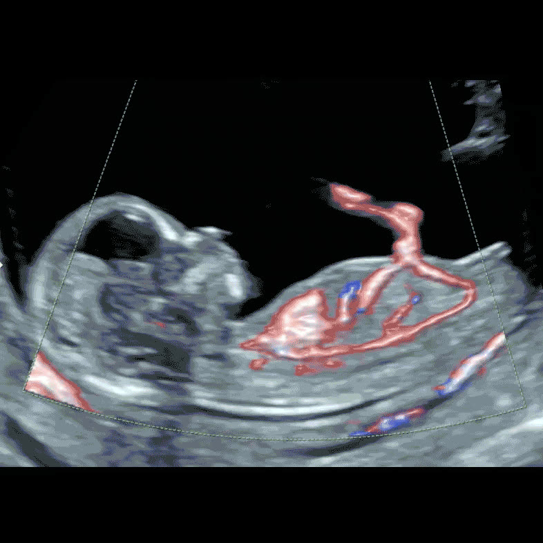
Colour Doppler image of fetal heart circulation at 13 weeks
Colour Doppler shows blood crossing the septum — often how a small VSD is first seen

VSD in numbers

Every figure below has a stated denominator and a named source. Where studies disagree we give the range, not the flattering end.

3 in 1,000 About 3 in every 1,000 babies are born with a ventricular septal defect — more than any other congenital heart defect (Liu et al., 2019)
~1 in 3 VSD makes up roughly a third of all congenital heart defects, which together affect about 9 in every 1,000 births (Liu et al., 2019)
86.9% Of muscular VSDs found by scanning more than 25,000 newborn babies, 86.9% had closed on their own by the first birthday (Copenhagen Baby Heart Study, 2024)
46.9% Perimembranous VSDs close less often: 46.9% had closed by one year in the same study (Copenhagen Baby Heart Study, 2024)
45.4% Of 55 babies whose isolated perimembranous VSD was found before birth, 45.4% closed in the womb (Gordin Kopylov et al., 2022)
5.5% In that same group of 55, only 3 babies (5.5%) needed heart surgery — all had holes larger than 3 mm (Gordin Kopylov et al., 2022)
99.4% 30-day survival after surgical closure of a VSD across UK and Irish centres, 2000–2016 (Farooqi et al., NCHDA, 2019)
96.9% Survival at 10 years for children treated for VSD in England and Wales, 2000–2022 (Brown et al., NCHDA, 2024)

Will the hole close on its own?

Very often, yes. The septum keeps growing after a VSD is found, and small holes get progressively walled off as the muscle thickens around them. The clearest evidence comes from the Copenhagen Baby Heart Study, which scanned the hearts of more than 25,000 newborn babies rather than waiting for a murmur to appear. It found a VSD in 3.

3% of newborns — far more than anyone counts clinically, because most are tiny. By the first birthday 83.5% had closed by themselves, and the proportion of one-year-olds with a VSD had fallen to 0.5% (Copenhagen Baby Heart Study, 2024). More than half of the closures happened within the first six months (Copenhagen Baby Heart Study, 2020).

Where the hole sits matters more than almost anything else. Muscular VSDs — holes in the thick lower part of the septum — closed on their own in 86.9% of cases within that first year. Perimembranous VSDs, which sit higher up near the valves, closed in 46.9% of cases. Subarterial defects, the rarest position, did not close at all during the first year (Copenhagen Baby Heart Study, 2024; 2020).

Closure can also happen before birth. In a series of 55 babies whose isolated perimembranous VSD was picked up on a fetal scan, the hole had already closed in the womb in 45.4% of cases, and more than half of the remainder closed during the first year. Only three babies — 5.5% — needed surgery, and all three had holes larger than 3 mm with symptoms of heart failure (Gordin Kopylov et al., 2022). Great Ormond Street Hospital tells parents the same in plain words: small VSDs seen on a prenatal scan often close without treatment, and may already be closed by the time the baby is born (GOSH).

Even when a small hole stays open, it is usually left alone rather than treated. Royal Brompton Hospital reports that around 70% of small VSDs get smaller as the child grows — a process that can take from a few months to 25 years — and that children with a small VSD have no symptoms, need no treatment, and can lead normal lives with unrestricted activity, including all sports (Royal Brompton, NHS).

Book fetal echo £360
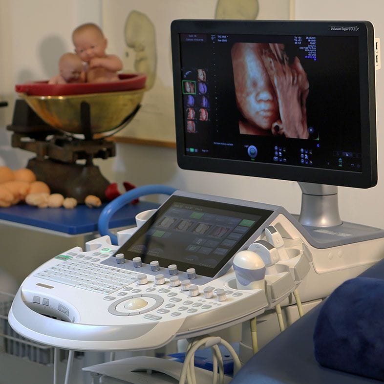
Ultrasound machine used for detailed fetal cardiac assessment
Small holes are often only visible with colour Doppler

Where the hole sits changes what happens next

Sonographers describe a VSD by its position in the septum, and that description is the best single predictor of what the next year will look like. If you have a report in front of you, the word you are looking for is usually one of these.

Best odds of closing

Muscular VSD

A hole in the thick, lower, muscular part of the septum. This is the type most often picked up when babies are scanned systematically, and by far the most likely to close by itself — 86.9% had closed within the first year in the Copenhagen Baby Heart Study (2024). Small muscular VSDs typically cause no symptoms, need no medication and no procedure, and are followed with a scan or two rather than treated. Many close before birth.

Perimembranous VSD

A hole in the thinner membranous part of the septum, higher up and close to the valves. It is the type most often diagnosed in children overall, and closes spontaneously less often than a muscular VSD — 46.9% within the first year in the Copenhagen data (2024). Even so, most found before birth are small and never need anything done: in the prenatal series of 55 isolated perimembranous VSDs, 45.4% closed in the womb and only 5.5% went on to surgery (Gordin Kopylov et al., 2022).

Inlet VSD

A hole low in the septum, just beneath the tricuspid and mitral valves — the inlet or atrioventricular canal region. Inlet defects are a small minority of VSDs and are seen more frequently in babies with trisomy 21 (StatPearls, 2024). Because the same part of the heart is involved in atrioventricular septal defect, this position prompts a careful look at the two valves and the crux of the heart, and a conversation about chromosomal testing.

Outlet VSD

A hole near the aortic and pulmonary valves, sometimes called subarterial or doubly committed. This is the exception to the reassuring picture above: outlet defects have low spontaneous closure rates (StatPearls, 2024), and in the Copenhagen Baby Heart Study not one subarterial VSD closed by itself during the first year (2020).

It is followed closely for a specific reason: sitting directly beneath the aortic valve, it leaves the right and non-coronary cusps without proper support, so aortic valve prolapse and leakage develop over time in a proportion of children (StatPearls, 2024).

That is what the follow-up echoes are watching for, and it is the usual reason an outlet VSD is repaired even when it is small.

Multiple VSDs

Several small holes rather than one, sometimes described as a ‘Swiss cheese’ septum. Individually they are often tiny and many still close on their own, but having more than one was among the factors associated with a lower chance of spontaneous closure in the Copenhagen Baby Heart Study (2024). These babies are followed for longer before anyone concludes the septum has finished closing.

Large VSD

The minority that genuinely needs treating. A large hole lets enough blood cross to the right side that the baby becomes breathless, sweaty during feeds and slow to gain weight, usually within the first weeks or months after birth. Treatment is medication first, then closure by open surgery or, in selected cases, a device passed through a catheter. Of the 55 prenatally diagnosed isolated perimembranous VSDs above, the three needing surgery were all larger than 3 mm with heart failure symptoms (Gordin Kopylov et al., 2022).

When a VSD is part of a bigger picture

This has to be said honestly, because it is why a VSD is worth a proper look rather than a shrug. A hole in the septum can occasionally be one visible component of something wider, and a specialist is checking for two things.

The first is other cardiac lesions. A VSD is a feature of several more complex conditions — tetralogy of Fallot, atrioventricular septal defect, coarctation of the aorta, double-outlet right ventricle. None of those is found by staring at the hole. They are found by working systematically through the outflow tracts, the great vessels, the arches and the valves, which is what a dedicated cardiac scan is for.

The second is a chromosomal or genetic condition. Where the VSD is genuinely isolated — nothing else abnormal anywhere on the scan — the chance of finding a chromosomal abnormality is low but not zero, and it would be wrong to give you a single tidy number for it.

Across the studies gathered in a 2024 review, the reported rate ranged from 0% to about 33%; that enormous spread reflects how strictly each study defined ‘isolated’, which test it used, and which families were referred in the first place, and the highest figures come from the older series (Frontiers in Genetics review, 2024).

Position appears to matter too: figures in that review put the rate at 3.13% for perimembranous VSDs against 0.93% for muscular ones, and Gómez and colleagues concluded that a muscular VSD carries a risk similar to that of a pregnancy with no finding at all. The most common single finding is a 22q11.

2 microdeletion, often called DiGeorge syndrome; a VSD is present in about 14% of people who have 22q11.2 deletion syndrome (Sauter et al., 2025). Heart differences are also common in Down’s syndrome — about half of all children with Down’s syndrome are born with a heart condition (Down’s Syndrome Association) — and in Edwards’ syndrome.

The asymmetry is what matters. A VSD found alongside another abnormality carries a considerably higher chance of an underlying genetic cause than one found entirely on its own.

In that series of 55 prenatally diagnosed isolated perimembranous VSDs, 34 families went on to genetic testing — 4 by NIPT and 30 by amniocentesis with a chromosomal microarray — and none of the 34 had a pathogenic or likely pathogenic finding (Gordin Kopylov et al., 2022).

Across babies found to have a heart condition of any kind, Royal Brompton puts the figure at about one in ten who will also have a problem affecting other organs or genes (Royal Brompton, NHS).

So a conversation about testing is worth having, and it is a conversation rather than an obligation. NIPT screens for the common trisomies from around 10 weeks, and some laboratories offer a 22q11.2 option — but it is a screening test, and a normal result does not exclude a microdeletion. Amniocentesis with a chromosomal microarray is the diagnostic test that can. Our genetic counselling service exists so that nobody has to weigh those options off the back of a leaflet handed over in a corridor.

What a fetal echo adds over a standard anomaly scan

A fetal echocardiogram is not a better version of the 20-week anomaly scan — it is a different examination with a different job. The anomaly scan surveys the whole baby and includes a defined set of heart views; a fetal echo does nothing but the heart. Here is what that buys you, and what it honestly cannot do.

Timing: when it is done

Most detailed fetal heart scans are performed between 18 and 23 weeks, when the structures are large enough to assess properly. In specialist centres a cardiac scan can be attempted from 13–14 weeks, early enough to answer some questions sooner, though not all (Royal Brompton, NHS). Our early fetal echocardiography covers that earlier window.

What the standard scan already covers

The NHS 20-week screening scan is not a cursory glance at the heart. It requires five specific cardiac views: situs and laterality, the four-chamber view, the left ventricular outflow tract, the right ventricular outflow tract or three-vessel view, and the three-vessel and trachea view (NHS Fetal Anomaly Screening Programme). Many VSDs are first spotted in exactly this examination.

What the echo adds

Time, magnification, and a systematic sweep of everything the hole might be attached to — valve function, the arches, the venous connections, and the direction and speed of flow across the defect on colour and pulsed Doppler. Small holes are often invisible on grey-scale imaging and appear only as a jet of colour, so a scan set up for cardiac Doppler finds things a survey scan is not designed to find.

What a fetal echo cannot do

It cannot promise a normal heart. Fetal heart scanning detects most structural abnormalities and most rhythm disturbances, but some are not visible before birth or only become evident afterwards, and atrial septal defect and patent ductus arteriosus cannot be diagnosed prenatally at all (Royal Brompton, NHS). A scan changes what is known — it does not change the heart.

Your questions answered

Is a VSD serious?

Usually not. VSD is the most common congenital heart defect — around 3 in every 1,000 births (Liu et al., 2019) — and the great majority of the holes found on scans are small. A small VSD usually causes no signs or symptoms at all, needs no treatment, and is compatible with a completely normal life (BHF).

The word ‘defect’ does a lot of unhelpful work here. It is a description of anatomy, not a prediction. Where a VSD is serious it is because the hole is large enough to overload the lungs, or because it is one part of a more complex heart condition — and both of those are things a detailed cardiac scan is designed to identify. The size and the position on your report tell you far more than the diagnosis on its own.

Will my baby need surgery?

Most babies with an isolated small VSD never have surgery. In a series of 55 babies whose isolated perimembranous VSD was found before birth, three needed an operation — 5.5% of the group — and all three had holes larger than 3 mm together with symptoms of heart failure (Gordin Kopylov et al., 2022). Across the prenatal series gathered in a 2024 review the proportion going to surgery ranged from 0% to about 20%, depending largely on how each study selected its cases and how big the defects were (Frontiers in Genetics review, 2024).

Where closure is needed, the UK results are strong. Across UK and Irish centres between 2000 and 2016, 30-day survival after surgical closure of a VSD was 99.4% (5,588 of 5,623 procedures); closure by a device passed through a catheter had a 30-day survival of 95.7% in the same audit (Farooqi et al., NCHDA, 2019).

Looking further out, among children in England and Wales who had a procedure for VSD between 2000 and 2022, survival was 98.4% at one year, 97.3% at five years and 96.9% at ten years (Brown et al., NCHDA, 2024). Those figures cover only children who had surgery or a catheter procedure, and include babies whose VSD was part of a more complex picture, not only the isolated ones.

Can a VSD close before my baby is born?

Yes, and it happens more often than most people expect. In the prenatal series of isolated perimembranous VSDs, 45.4% had closed in the womb — some before 30 weeks, some between 30 weeks and the first month of life (Gordin Kopylov et al., 2022). Great Ormond Street Hospital tells parents that small VSDs seen on a prenatal scan often close without treatment and may already be closed by the time the baby is born (GOSH).

This is why a repeat scan later in pregnancy is often suggested rather than a plan for treatment. It is not a delaying tactic — for a meaningful proportion of babies, the finding simply resolves itself, and the follow-up scan is the thing that confirms it.

What causes a hole in a baby’s heart?

In most cases nothing that anybody did or failed to do. The septum forms during the first eight weeks of pregnancy, and a VSD is what happens when the two growing walls do not quite meet. That process is largely complete before many women even know they are pregnant.

Some VSDs sit within a wider genetic picture, and known risk factors for congenital heart defects generally include maternal diabetes, rubella infection in pregnancy, certain medications, and smoking or alcohol — these are covered on our congenital heart defects page. But for the ordinary small muscular hole found at a scan, there is no identifiable cause and no behaviour to trace back to. It is worth saying that plainly, because a great many parents spend the drive home doing exactly that.

Does a VSD mean my baby has Down’s syndrome?

No — but it is a fair question, and the honest answer is that the published numbers are all over the place.

Where the VSD is genuinely isolated, with nothing else abnormal on the scan, the reported chance of finding a chromosomal abnormality ranges from 0% to about 33% across the studies gathered in a 2024 review, depending heavily on how each study defined ‘isolated’, which test it used and which families were referred; the highest figures come from the older series (Frontiers in Genetics review, 2024).

The most directly relevant modern evidence is more reassuring: in a series of 55 prenatally diagnosed isolated perimembranous VSDs, none of the 34 fetuses tested had a pathogenic or likely pathogenic finding (Gordin Kopylov et al., 2022). But a genuinely isolated VSD is not a zero-risk finding, and nobody should tell you it is.

The connection runs more strongly in the other direction: about half of all children with Down’s syndrome are born with a heart condition (Down’s Syndrome Association), and heart differences are also common in Edwards’ syndrome. So a heart finding is a reason to review the rest of the scan carefully and to discuss testing — not a reason to conclude anything. Where the VSD sits low in the septum, in the inlet position, that association with trisomy 21 is somewhat stronger (StatPearls, 2024).

Should I have NIPT or an amniocentesis if a VSD has been found?

It depends on what else the scan showed, and it is genuinely your decision. NIPT is a screening test from a maternal blood sample, available from around 10 weeks, and screens for the common trisomies; some laboratories also offer a 22q11.2 microdeletion option. It carries no risk to the pregnancy, but a normal NIPT result cannot exclude a microdeletion or a smaller genetic change.

Amniocentesis with a chromosomal microarray is the diagnostic test that can. It is a more definitive answer and carries a small procedural risk. Which of these is proportionate depends on whether the VSD is isolated, its size and position, and what you would do with the information — which is exactly what genetic counselling is for. In the prenatal series of 55 isolated perimembranous VSDs, 34 families went on to testing — 4 by NIPT and 30 by amniocentesis with a microarray — and none had a pathogenic or likely pathogenic finding (Gordin Kopylov et al., 2022).

How big does a VSD have to be to matter?

There is no single threshold, but size is the most informative number on your report. In the prenatal series, every baby who eventually needed surgery had a defect larger than 3 mm, and all of them had symptoms of heart failure after birth; the smaller holes did not need an operation (Gordin Kopylov et al., 2022). Small size was also one of the factors most strongly associated with spontaneous closure in the Copenhagen Baby Heart Study (2020).

Size is judged relative to the baby, not in absolute millimetres alone, and it changes as the heart grows — a hole that is unchanged in millimetres is proportionally smaller at 34 weeks than it was at 20. This is one of the main reasons a follow-up scan is useful.

What happens after my baby is born?

For an isolated small VSD, usually very little. Great Ormond Street advises that the baby should be well, with no concerning symptoms; if the hole is still open, doctors will hear a murmur, and an echocardiogram with a cardiologist or paediatrician is arranged in the first few weeks of life (GOSH). A murmur in this situation is not a bad sign — it is the sound of a small hole, and a loud murmur often means a small defect rather than a large one.

Where the hole is large, the picture declares itself in the first weeks: breathlessness, sweating during feeds, and slow weight gain. That is managed with medication first and closure afterwards, either surgically or, in selected cases, with a device delivered through a catheter (BHF). Either way, delivery does not usually need to happen anywhere unusual for an isolated small VSD — that is a decision your team will make on the whole picture.

Will my baby be able to feed and grow normally?

With a small VSD, yes. Royal Brompton states that patients with a small VSD have no symptoms and need no treatment, and that children with small VSDs can expect to lead normal lives (Royal Brompton, NHS). Nothing about a small hole limits what your baby can do — though the same guidance asks that your baby be weighed once or twice a month at first, so that growth is checked rather than assumed.

Feeding difficulty is the main thing paediatric teams watch for, because it is the earliest practical sign that a hole is large enough to matter: a baby who tires or sweats partway through a feed, and whose weight is drifting downwards on the centile chart, is the baby who gets reviewed sooner. If your baby feeds well and gains weight, that is meaningful reassurance in itself.

Can a scan miss a VSD, or say there is one when there isn’t?

Both are possible, and it would be dishonest to say otherwise. Small muscular VSDs are often invisible on grey-scale ultrasound and show up only as a jet of colour on Doppler, so whether one is seen depends on the equipment, the baby’s position, and how long is spent on the heart. Fetal heart scanning detects most structural abnormalities and rhythm disturbances, but some are not visible before birth or only become evident afterwards, and atrial septal defect and patent ductus arteriosus cannot be diagnosed prenatally at all (Royal Brompton, NHS).

The reverse also happens: an apparent gap in the septum can be an artefact of the angle the heart is being viewed from, which is why a suspected VSD is confirmed from more than one plane before it is reported. If a finding is uncertain, a repeat or a second opinion scan is a reasonable next step rather than a sign that something has gone wrong.

When is a fetal echo done, and what does it involve?

Most detailed fetal heart scans are performed between 18 and 23 weeks, when the cardiac structures are big enough to assess reliably. In specialist centres a cardiac scan can be attempted from 13–14 weeks (Royal Brompton, NHS), which is the window our early fetal echocardiography service covers.

The examination itself is an ordinary abdominal ultrasound — no needles, nothing given to you or the baby — but it is longer and narrower in focus than a routine scan. The chambers, valves, outflow tracts, great vessels and arches are each assessed in turn, with colour and pulsed Doppler used to look at how blood is moving, including across the defect. You will be told what is seen during the scan rather than afterwards. Our fetal echocardiography appointment is £360.

Will my child be able to play sport and live normally?

For a small VSD, yes. Royal Brompton states it directly: all children with small VSDs can expect to lead normal lives, their activities can be unrestricted, and they are able to take part in all sports (Royal Brompton, NHS). The British Heart Foundation says the same of repaired defects — most people with a small VSD or a repaired VSD live a normal, healthy life (BHF).

Follow-up with a cardiologist usually continues while the hole remains open, and good dental hygiene is emphasised because it reduces the risk of infection reaching the heart. Neither of those is a restriction on ordinary life. If you are reading this in a hospital car park, this is the paragraph worth taking away.

Contact

Speak to us about a detailed look at your baby’s heart

Send us an enquiry

A clinician will reply within one working day.

For urgent matters please call 020 3687 2939.

Sources & clinical references

The figures and clinical statements on this page are drawn from the sources below. Guidance evolves — always discuss your individual circumstances with a clinician.

  1. Royal Brompton & Harefield hospitals (Guy’s and St Thomas’ NHS Foundation Trust)Small ventricular septal defect (VSD)2024
  2. Royal Brompton & Harefield hospitals (Guy’s and St Thomas’ NHS Foundation Trust)Fetal cardiology scan2024
  3. Great Ormond Street Hospital for Children NHS Foundation TrustPrenatal ultrasound scans showing a small ventricular septal defect2024
  4. British Heart FoundationVentricular septal defect (VSD)2024
  5. NHS Fetal Anomaly Screening Programme (GOV.UK)Fetal anomaly screening programme handbook: 20-week screening scan2023
  6. International Journal of Epidemiology (Liu et al.)Global birth prevalence of congenital heart defects 1970–2017: updated systematic review and meta-analysis of 260 studies2019
  7. Neonatology (Copenhagen Baby Heart Study)The prevalence and spontaneous closure of ventricular septal defects the first year of life2024
  8. European Heart Journal (Copenhagen Baby Heart Study, congress abstract)Spontaneous closure of ventricular septal defects in newborns the first year of life2020
  9. Prenatal Diagnosis (Gordin Kopylov et al.)Prenatally diagnosed isolated perimembranous ventricular septal defect: genetic and clinical implications2022
  10. Heart (Farooqi et al.) — National Congenital Heart Disease Audit dataTrends in surgical and catheter interventions for isolated congenital shunt lesions in the UK and Ireland2019
  11. Journal of the American Heart Association (Brown et al.) — National Congenital Heart Disease Audit, England and WalesEvaluating long-term outcomes of children undergoing surgical treatment for congenital heart disease for national audit in England and Wales2024
  12. Frontiers in GeneticsPrenatal finding of isolated ventricular septal defect: genetic association, outcomes and counseling2024
  13. Journal of Medical Genetics (Sauter et al.)Congenital heart disease in 22q11.2 deletion syndrome: a meta-analysis and systematic review of the literature2025
  14. StatPearls (NCBI Bookshelf)Ventricular Septal Defect2024
  15. Down’s Syndrome Association (UK)The heart and circulation2024