Oligohydramnios

Oligohydramnios means there is less fluid around your baby than expected — the pool measured on the scan is shallower than the chart says it should be. It is a finding, not a diagnosis, and the useful question is always why. Near or past your due date, low fluid on its own, in a well-grown baby who is moving normally, most often ends with a normal birth of a healthy baby. It matters more when the membranes may have ruptured, when the placenta is not keeping up — which is why it travels with fetal growth restriction — or when the fluid is very low early in pregnancy.

Available atLPC City (E1)LPC West (SW10)

What low amniotic fluid actually means

Amniotic fluid is mostly your baby's urine. From around 16 weeks the kidneys make it and your baby swallows it, and the pool seen on a scan is the balance between the two. So a low reading is telling you something about that loop — the membranes holding it in, the placenta supplying the blood the kidneys filter, or the kidneys and bladder themselves. That is the whole logic behind the investigations that follow.

Volume is not fixed. It rises through pregnancy, peaks in the third trimester and then falls, so a pocket that would worry at 28 weeks can be unremarkable at 41. Oligohydramnios affects under 1% of preterm pregnancies and becomes steadily more common past the due date: about 1.1% between 37 and 39 weeks, 1.7% at 40, 2.7% at 41 and 4.4% by 42 in a Norwegian cohort.

What changes the picture is context — whether your waters may have broken, whether your baby is measuring small, and what the blood flow shows on a growth scan. A fluid figure read on its own has caused a great deal of unnecessary alarm, and a fair amount of unnecessary intervention.

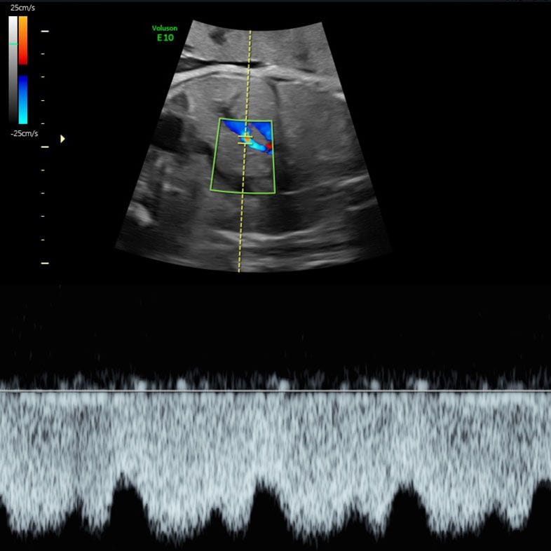
Third-trimester ultrasound image showing a baby and the fluid around them

If your waters may have broken, phone your maternity unit today

Ruptured membranes are the commonest obstetric cause of oligohydramnios, and the most time-critical — preterm prelabour rupture alone accounts for more than 37% of cases found in the second and third trimesters. You do not always get a dramatic gush: a persistent trickle, or feeling damp without explanation, counts. The NHS advises calling your midwife or maternity unit about a gush or trickle of fluid, and calling immediately if your baby is moving less or differently — not waiting until the next day, even in the middle of the night. Rupture is diagnosed by a sterile speculum examination, not by ultrasound, so a private scan is the wrong first step.

Oligohydramnios in numbers

Under 1% Oligohydramnios affects fewer than 1% of preterm pregnancies (StatPearls, 2024)
1.1% → 4.4% Prevalence rises from about 1.1% at 37–39 weeks to 4.4% by 42 weeks in a Norwegian cohort (StatPearls, 2024)
More than 1 in 3 Preterm prelabour rupture of membranes alone accounts for over 37% of oligohydramnios found in the second and third trimesters (StatPearls, 2024)
Up to 3% Preterm prelabour rupture of membranes complicates up to 3% of pregnancies and precedes 30–40% of preterm births (RCOG, 2019)
2.39× AFI diagnoses oligohydramnios 2.39 times as often as the deepest-pocket method, across 5 trials and 3,226 women (Cochrane, 2008)
No clear difference In 679 term cases of isolated low fluid versus 3,264 with normal fluid, neonatal outcomes did not differ (Rossi & Prefumo, 2013). A larger review of 35,999 women did find small increases in Apgar below 7 (OR 2.01) and neonatal unit admission (OR 1.47) (Shrem et al., 2016)

How the fluid is measured — and why the method matters

Amniotic fluid cannot be measured directly without a dye study, so ultrasound estimates it one of two ways. Which one your unit uses genuinely changes how likely you are to be told you have oligohydramnios — worth knowing before a number frightens you.

Single deepest pocket (SDP)

The sonographer finds the largest clear pool of fluid, free of cord and limbs, and measures its depth. Normal is 2 cm or more and under 8 cm; below 2 cm is oligohydramnios. One measurement, quickly taken, and the method most UK units now favour.

Amniotic fluid index (AFI)

The uterus is divided into four quadrants, the deepest pocket in each is measured, and the four are added together. Normal is above 5 cm and below 24 cm; 5 cm or less is oligohydramnios. Four measurements mean four chances for error, and it flags far more pregnancies than the deepest pocket does.

Why the method changes the answer

In a Cochrane review of five randomised trials and 3,226 women, using AFI rather than the deepest pocket diagnosed oligohydramnios 2.39 times as often (RR 2.39, 95% CI 1.73 to 3.28), nearly doubled inductions of labour (RR 1.92) and increased caesareans for fetal distress (RR 1.46) — with no difference in neonatal unit admission (RR 1.04), cord pH, meconium or Apgar score. More diagnoses, more intervention, no measurable benefit to babies. Ask which method your scan used.

What the measurement cannot tell you

Fluid volume is one line in a report. It does not say whether your baby is growing well, whether the placenta is coping, or whether the membranes are intact. It also moves with your baby's position and bladder, and with how the probe is angled and pressed — so a single borderline figure in a well baby is normally repeated rather than acted on.

Why the fluid might be low

Ruptured membranes — checked first

The commonest cause and the most urgent. If the sac has leaked, fluid falls because it is escaping, and before 37 weeks that carries real risks of infection, cord problems and preterm birth. Preterm prelabour rupture complicates up to 3% of pregnancies and precedes 30–40% of preterm births (RCOG, 2019). Diagnosis is a sterile speculum examination, sometimes with a swab test — not a scan.

Placental insufficiency

If the placenta is not delivering enough blood, your baby diverts what there is to the brain and heart, the kidneys receive less, and less urine is made. That is why low fluid so often sits alongside fetal growth restriction and conditions such as pre-eclampsia. It is also why the response is rarely another fluid measurement: the growth centiles and the umbilical artery Doppler show whether the placenta is the problem.

Your baby's kidneys or urinary tract

Because the fluid is mostly fetal urine, a kidney or bladder problem shows up as low fluid — and very low fluid in the second trimester points here first. Bilateral renal agenesis, where neither kidney forms, leaves very little or no fluid by the 20-week scan; it affects about 1 in 5,000 babies and is not survivable, because lungs need fluid to develop. Blockages lower in the urinary tract are far less severe and often treatable, and anatomy is reviewed on the anomaly scan.

Going past your due date

Fluid falls naturally in late pregnancy. Prevalence climbs from roughly 1.1% between 37 and 39 weeks to 2.7% at 41 and 4.4% by 42. The NHS offers induction if labour has not started by 41 weeks, and increased monitoring beyond 42 weeks if you decline. At this stage low fluid is usually one input into a conversation about timing rather than a new problem.

Medication

Anti-inflammatory painkillers reduce blood flow through your baby's kidneys. The MHRA advises avoiding systemic NSAIDs — ibuprofen, naproxen, diclofenac — from week 20 unless clinically required, at the lowest dose for the shortest time, because prolonged use can cause oligohydramnios through fetal renal dysfunction. It can appear soon after starting and is usually reversible on stopping. ACE inhibitors and angiotensin receptor blockers act similarly. Tell your midwife what you have taken.

Why it matters — and how much

The honest answer depends almost entirely on whether the low fluid is isolated — membranes intact, growth normal, Dopplers normal, nothing else going on. In that situation the published evidence is genuinely reassuring, and it is worth seeing the actual numbers rather than being told simply not to worry.

A meta-analysis of four studies covering 679 term and post-term pregnancies with isolated oligohydramnios, against 3,264 with normal fluid, found more obstetric intervention — caesarean or operative delivery for a non-reassuring heart rate, odds ratio 2.30 (95% CI 1.00 to 5.

29) — but no significant difference in meconium, Apgar below 7 at five minutes, cord pH, neonatal unit admission, babies born small, or perinatal death (Rossi & Prefumo, 2013). A larger review of 12 studies and 35,999 women, 2,414 of them with isolated oligohydramnios, found the same pattern more strongly: induction odds ratio 7.56 and caesarean 2.

07, with modest increases in Apgar below 7 (OR 2.01) and neonatal unit admission (OR 1.47), and no difference in cord pH or meconium (Shrem et al., 2016).

Read together, those studies say something specific: isolated low fluid at term changes what happens to you considerably more than what happens to your baby. Where it genuinely matters is when it is not isolated — alongside a faltering growth trend, an abnormal umbilical or middle cerebral artery Doppler, or reduced movements. NHS England's Saving Babies' Lives care bundle lists reduced liquor volume among the findings that should trigger a review of the timing of birth, precisely because of the company it keeps. That is a reason for closer monitoring, not a prediction.

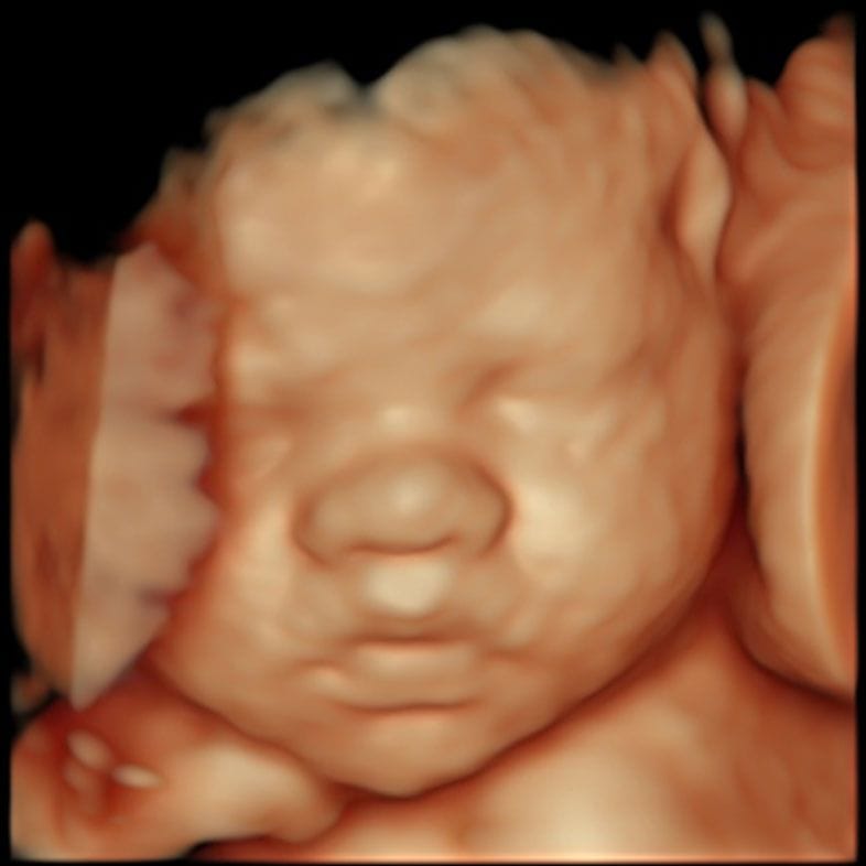
Doppler waveform showing blood flow in a fetal vessel during a third-trimester scan
Dopplers, not fluid volume alone, show whether the placenta is keeping up

What usually happens next

1

Ruling out ruptured membranes

A sterile speculum examination, and where no fluid is seen, an IGFBP-1 or PAMG-1 swab. This comes first because it changes everything after it: before 37 weeks, confirmed rupture means antibiotics, steroids where appropriate, and close surveillance.

2

Re-measuring the fluid

The pocket is measured again, ideally by the deepest-pocket method, with your baby's bladder and stomach checked to confirm urine is being passed and fluid swallowed.

3

Assessing growth and blood flow

Head, abdomen and thigh bone are measured for an estimated weight and plotted as a centile, and the umbilical and middle cerebral artery Dopplers recorded. This separates low fluid that reflects a struggling placenta from low fluid that does not.

4

Looking for a cause

The kidneys, bladder and remaining anatomy are reviewed, your medications checked, and your blood pressure and urine tested for pre-eclampsia. Where the fluid is very low and the pregnancy early, referral to a fetal medicine unit follows.

5

Agreeing a monitoring plan

If everything else is normal, the usual answer is repeat scanning and a conversation about timing rather than immediate action. If growth or Doppler findings are abnormal, monitoring intensifies and the timing of birth is reviewed with your obstetric team.

What a scan can and cannot tell you

A private wellbeing scan or growth scan can measure the fluid, the growth and the Dopplers on the day and give you a written report your NHS team can use. It cannot change how your placenta works, and it is not the right first step if you think you may be leaking.

  • It can measure both the deepest pocket and the amniotic fluid index, and state which was used — useful, because the two disagree often.
  • It can estimate your baby's weight and plot it on a centile chart, so a small measurement and a low fluid reading are seen together.
  • It can record umbilical and middle cerebral artery Dopplers, which is where a struggling placenta shows itself before size does.
  • It can check the kidneys, the bladder filling and emptying, and the rest of the anatomy for a structural cause.
  • It cannot diagnose ruptured membranes. That needs a speculum examination at your maternity unit, and it should not wait.
  • It cannot treat low fluid, and no scan changes an outcome by being performed.

Your questions answered

What does oligohydramnios mean?

It means there is less amniotic fluid around your baby than expected. On ultrasound that is a single deepest pocket under 2 cm, or an amniotic fluid index of 5 cm or less. It is a measurement rather than a diagnosis: it tells your team to look for a reason. Near or past your due date, the reason is often nothing more than the natural fall in fluid at the end of pregnancy.

Is low amniotic fluid dangerous for my baby?

It depends on why it is low. Where it is isolated at term — membranes intact, growth normal, Dopplers normal — pooled evidence from four studies and 679 cases shows more obstetric intervention — caesarean or operative delivery for a non-reassuring heart rate — but neonatal outcomes broadly the same as pregnancies with normal fluid (Rossi & Prefumo, 2013). Alongside restricted growth, abnormal blood flow or ruptured membranes it matters much more. Very low fluid early in pregnancy is the real concern, because lungs need fluid to develop.

What is a normal amniotic fluid index?

An AFI above 5 cm and below 24 cm is normal; 5 cm or less is oligohydramnios. If your unit uses the single deepest pocket instead, normal is 2 cm up to 8 cm, and under 2 cm is low. Ask which method was used — the numbers are not interchangeable, and the same pregnancy can be labelled differently by each.

Why did one scan say my fluid was low and the next one said it was fine?

Because measuring fluid is imprecise. The pocket depends on how your baby is lying, how full their bladder is, and how the probe is angled and pressed, and the volume itself shifts over hours. It is also why the amniotic fluid index, which adds four measurements together, labels 2.39 times as many pregnancies as the deepest pocket does (Cochrane, 2008). A single low reading in a well baby is usually repeated before anything is decided.

Can low amniotic fluid mean my waters have broken?

Yes, and it is the first thing to rule out — preterm prelabour rupture alone accounts for more than 37% of oligohydramnios found in the second and third trimesters. It does not always announce itself with a gush; a persistent trickle, or feeling damp with no explanation, is enough. It is diagnosed by a sterile speculum examination at your maternity unit, not by ultrasound. If there is any possibility, phone them today.

Can drinking more water increase my amniotic fluid?

It raises the measured number. A Cochrane review of four trials and 122 women found that drinking around two litres of water increased the amniotic fluid index in oligohydramnios by a mean difference of 2.01 (95% CI 1.43 to 2.60) — but no clinically important outcomes were assessed in any of the trials. Hydration changes the measurement; it does not repair ruptured membranes or a struggling placenta.

Does low fluid mean I will be induced or need a caesarean?

It makes both more likely. In a review of 12 studies and 35,999 women, isolated oligohydramnios at term raised the odds of induction more than sevenfold (OR 7.56, 95% CI 4.58 to 12.48) and roughly doubled caesareans (OR 2.07, 95% CI 1.77 to 2.41) (Shrem et al., 2016). Whether that intervention helps is less settled: in the single randomised trial of induction versus expectant management, no significant outcome differed. It is worth discussing rather than accepting automatically.

Does oligohydramnios mean my baby has kidney problems?

Usually not, particularly at term. But because the fluid is mostly your baby's urine, kidney and urinary tract problems are the first thing looked for when fluid is very low in the second trimester. Bilateral renal agenesis, where neither kidney forms, occurs in about 1 in 5,000 babies and leaves very little or no fluid by the 20-week scan. Far more often the kidneys and bladder look normal.

I am past my due date and they said my fluid is on the low side — should I be worried?

This is the most common version of oligohydramnios and the least alarming. Fluid falls naturally after 37 weeks: prevalence goes from around 1.1% between 37 and 39 weeks to 1.7% at 40, 2.7% at 41 and 4.4% by 42. It feeds into a conversation about induction, which the NHS offers if labour has not started by 41 weeks. On its own it is not evidence that your baby is unwell.

Can low amniotic fluid be treated?

Not in a way that removes the cause. Where a medicine is responsible, stopping it usually restores the fluid — the MHRA notes NSAID-related oligohydramnios is generally reversible on discontinuation. Amnioinfusion is used in specific situations rather than as a treatment for the pregnancy, and trials of serial amnioinfusion in mid-trimester rupture of membranes have not improved survival. Otherwise the response is monitoring and getting the timing of birth right.

What are the warning signs I should not ignore?

A gush or trickle of fluid from the vagina at any stage. Any change in your baby's movements — fewer, weaker, or a different pattern. Vaginal bleeding. Feeling feverish, hot, cold or shivery, or fluid that looks greenish or smells. Abdominal pain, or contractions before 37 weeks. Each means phoning your midwife or maternity unit straight away, not waiting for an appointment or a private scan. On movements the NHS is explicit: do not wait until the next day, call immediately, even in the middle of the night.

Will my baby be all right?

For most babies found to have low fluid at term with nothing else wrong, yes — and that is by far the largest group. No scan and no clinician can promise you an outcome, and you should be wary of anyone who does. What is true is that the situations where oligohydramnios genuinely threatens a baby are usually identifiable: ruptured membranes, restricted growth with abnormal Dopplers, or very low fluid in the second trimester. If none of those applies to you, the evidence is on your side. If your baby is moving less, contact your maternity unit the same day.

Contact

Speak to us about low amniotic fluid

Send us an enquiry

A clinician will reply within one working day.

For urgent matters please call 020 3687 2939.

Sources & clinical references

The figures and clinical statements on this page are drawn from the sources below. Guidance evolves — always discuss your individual circumstances with a clinician.

  1. NHSPremature labour and birth — waters breaking early2025
  2. NHSYour baby's movements2025
  3. Royal College of Obstetricians and Gynaecologists (RCOG)Care of Women Presenting with Suspected Preterm Prelabour Rupture of Membranes from 24+0 Weeks of Gestation (Green-top Guideline No. 73)2019
  4. Cochrane Database of Systematic Reviews (Nabhan & Abdelmoula)Amniotic fluid index compared with single deepest vertical pocket measurement in predicting an adverse pregnancy outcome2008
  5. StatPearls, NCBI BookshelfOligohydramnios2024
  6. European Journal of Obstetrics & Gynecology and Reproductive Biology (Rossi & Prefumo), via NCBI BookshelfPerinatal outcomes of isolated oligohydramnios at term and post-term pregnancy: a systematic review of literature with meta-analysis2013
  7. Fetal Diagnosis and Therapy (Shrem et al.), KargerIsolated Oligohydramnios at Term as an Indication for Labor Induction: A Systematic Review and Meta-Analysis2016
  8. Cochrane Database of Systematic Reviews (Hofmeyr, Gülmezoglu & Novikova)Maternal hydration for increasing amniotic fluid volume in oligohydramnios and normal amniotic fluid volume2002
  9. GOV.UK (Medicines and Healthcare products Regulatory Agency)Non-steroidal anti-inflammatory drugs (NSAIDs): potential risks following prolonged use after 20 weeks of pregnancy2023
  10. GOV.UK (NHS Fetal Anomaly Screening Programme)Bilateral renal agenesis (BRA): information for parents2026
  11. NHS EnglandSaving babies' lives version 3: a care bundle for reducing perinatal mortality2023
  12. NHSInducing labour2025