
Frank breech (extended)
Hips bent, both legs straight up in front of the body, feet somewhere near the face. This is the position most people picture when they hear the word breech. Cord prolapse is least likely here — reported at around 0.5%.
Breech simply means your baby is lying bottom or feet first rather than head down. Here is the fact most pages leave out: about one baby in four is breech at 28 weeks, roughly 7 in 100 at 32 weeks, and only 3–4 in 100 by the time labour starts. Almost all of them turn on their own, with nobody doing anything. Even when a baby is still breech at 36 weeks, a study of 107,875 pregnancies found 43.8% were head down by birth — and three-quarters of those turned by themselves. A presentation scan tells you which way your baby is lying. It does not turn them.
Available atLPC City (E1)LPC West (SW10)
Presentation is just a word for which part of your baby is closest to the way out. Head down — cephalic — is the commonest. Breech means bottom or feet first. Transverse or oblique means lying across the uterus or at an angle. None of these is a diagnosis of anything being wrong. It is a description of how your baby happens to be lying today.
Being breech in the second and early third trimester is a normal stage, not a problem waiting to be solved. Around 25% of babies are breech at 28 weeks or earlier, and 7% at 32 weeks; at term it is 3–4 in 100. The turn usually happens by itself somewhere between roughly 30 and 36 weeks, as a growing baby settles into the shape that fits best. Nobody has to make it happen, and there is nothing you did that caused it.
Some things make a baby more likely to stay breech: being born early, twins, a low-lying placenta, more or less amniotic fluid than usual, fibroids, or an unusually shaped uterus. Occasionally a baby who stays breech is a clue to something structural, which is why a scan looks at the anatomy as well as the position — see common fetal anomalies. Most of the time no cause is ever found, and that is by far the commonest answer.
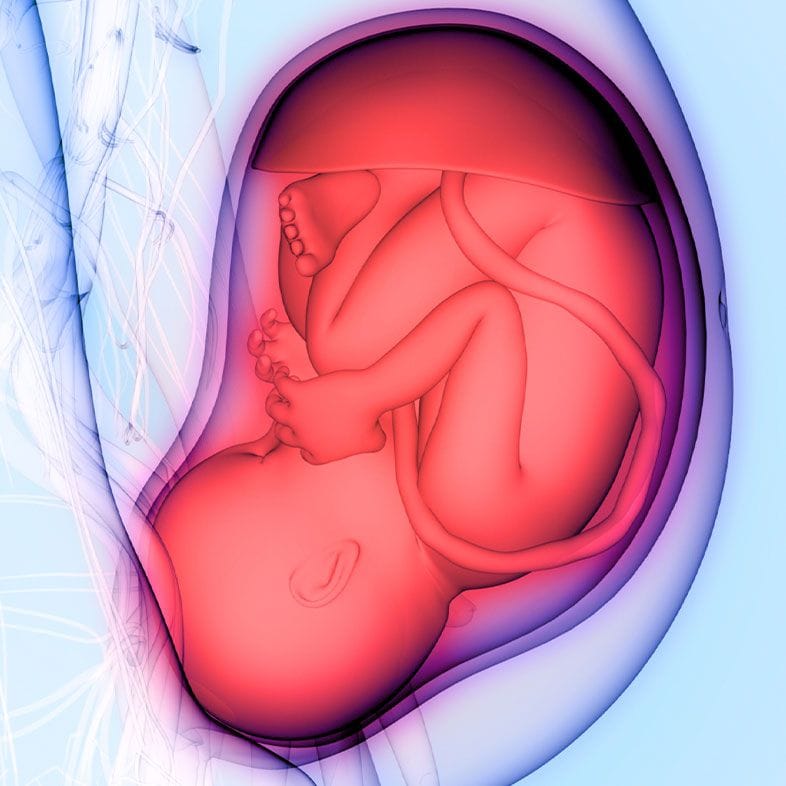
These describe populations, not a prediction for your pregnancy.
Breech is not one position. Which one your baby is in does not change how likely they are to turn, but it does change the birth conversation, so it is worth knowing the word your notes use.
Hips bent, both legs straight up in front of the body, feet somewhere near the face. This is the position most people picture when they hear the word breech. Cord prolapse is least likely here — reported at around 0.5%.
Your baby sits cross-legged, hips and knees both bent, bottom lowest. The reported risk of cord prolapse sits in the middle, at around 4–6%.
One or both feet, or a knee, lying lower than the bottom. This one matters more than the others: the reported risk of cord prolapse is 15–18%, and RCOG lists a footling presentation among the factors that make a planned vaginal breech birth higher risk.
You cannot work out which type you have by feel, and neither can anyone else without a scan — it takes one line on a report. It is worth asking about your baby's neck at the same time: a hyperextended neck on ultrasound is another of RCOG's higher-risk markers for a planned vaginal breech birth.
The first check is hands. A midwife feels the bump for the hard round head and the softer bottom. It is quick, free and a perfectly reasonable first pass — and it is also the weakest link in the chain. NICE's evidence review puts the sensitivity of abdominal palpation for breech at 57–70%, against essentially 100% sensitivity and specificity for ultrasound.
In the study behind the upper end of that range, 1,633 women were examined at 35–37 weeks and then scanned: palpation found 70% of the non-cephalic babies (95% CI 62–78%), and specificity was 95%. Among women with obesity it found only 3 of the 8 non-cephalic babies — 38% — though that is a small enough subgroup to be read as a signal rather than a precise figure.
That gap is why breech so often arrives as a surprise. In the Cambridge POP study, 3,879 first-time mothers were scanned at 36 weeks; 179 (4.6%) were breech, and in 96 of those cases — 53.6% — nobody had suspected it beforehand. In Oxfordshire, before a routine late scan was introduced, 22.3% of breech babies were not identified until labour had started. Afterwards it was 4.7% (RR 0.21, 95% CI 0.12–0.36), across 27,825 pregnancies.
What that does not prove is that scanning everyone makes babies safer, and it would be dishonest to imply otherwise. In the same Oxfordshire cohort the fall in emergency caesareans, from 32.4% to 27.8%, was not statistically significant, and the reductions in markers of newborn illness did not reach significance either. NICE's committee looked at the same evidence and kept selective scanning — scan when palpation raises a suspicion — rather than scanning every pregnancy. So ultrasound is the right way to answer the question is my baby breech. It is not, on current evidence, a way to buy a better outcome.
Cephalic (head down), breech (bottom or feet first), or transverse or oblique (across, or at an angle). This is the question ultrasound answers definitively, where hands answer it correctly 57–70% of the time.
Frank, complete or footling, and whether your baby's neck is flexed or hyperextended. A footling presentation and a hyperextended neck are both on RCOG's list of features that make a planned vaginal breech birth higher risk, so knowing which one you have changes the conversation you are about to have.
Head, abdomen and thigh bone combined into an estimate. RCOG treats an estimated weight above 3.8 kg, and one below the 10th centile, as markers of higher risk for planned vaginal breech birth. It is an estimate with a genuine margin of error, so it is read alongside everything else rather than on its own.
How much amniotic fluid there is, where the placenta is sitting and where the cord lies. These matter in their own right, and also because low fluid or a low-lying placenta can be the reason an ECV is not offered.
A written report on the day — position, type, measurements and images — in a form your midwife or obstetric team can act on. If you are arranging the rest of your care privately, see private maternity care. If your real question is how your baby is coping rather than which way round they are, a wellbeing scan is the closer fit.
It cannot turn your baby. It cannot promise you an outcome, and paying for it does not change one. It does not replace your NHS antenatal care or your maternity unit's plan. What it gives you is information sooner, which is worth something when the window for deciding is about a fortnight wide.
ECV — external cephalic version — is a doctor turning your baby by hand, from the outside, with firm steady pressure on your abdomen. It is done in a hospital obstetric unit rather than at a scanning clinic: your baby's heartbeat is monitored before and after, ultrasound checks the position and the fluid, and a medicine to relax the uterus is often given first. The attempt itself takes a few minutes. It is usually uncomfortable rather than painful, and you can ask for it to stop at any point.
RCOG's position is that women with a breech baby at term should be offered ECV unless there is an absolute reason not to. It is usually done after 36 or 37 weeks — late enough that a baby who turns is unlikely to flip back, early enough to leave the birth options open — and it can be attempted right up to the early stages of labour.
It works about half the time. RCOG quotes roughly 50%. In a 2025 series from King's College Hospital and Medway Maritime Hospital, 698 of 1,584 attempts succeeded — 44.1% — and RCOG says it is more likely to work if you have given birth vaginally before. Pooled randomised trials (8 trials, 1,305 women) found that attempting ECV reduced non-cephalic presentation at birth (RR 0.42, 95% CI 0.29–0.61) and caesarean birth (RR 0.57, 95% CI 0.40–0.82), though Cochrane graded the certainty of that evidence as low to very low.
The risk figure worth carrying into the conversation is this: about 1 in 200 women need an emergency caesarean immediately after an ECV, because of bleeding from the placenta or a change in the baby's heartbeat. That is precisely why it is done on a labour ward with monitoring rather than in a clinic. RCOG describes ECV overall as generally safe with a very low complication rate, and says that overall there does not appear to be an increased risk to your baby.
Not everyone has one, and that is normal. In that same series, ECV was attempted in only 1,584 of 6,211 non-cephalic pregnancies — 25.5%. Some women declined it; some had a reason it could not be done, such as low fluid or a low-lying placenta; some were already booked for a caesarean for a separate reason; and some babies turned on their own before the appointment came round. Declining an ECV is a legitimate choice, not a failure to comply.
RCOG is unusually direct about how this conversation should go: clinicians should counsel women in an unbiased way that ensures a proper understanding of the absolute as well as the relative risks. So here are the absolute numbers. The decision belongs to you and your obstetric team, and this page is not going to steer it.
| What the evidence shows | Planned caesarean | Planned vaginal breech birth |
|---|---|---|
| Baby dying around the time of birth (RCOG) | About 0.5 in 1,000, when planned after 39+0 weeks | About 2 in 1,000 |
| Ends in an emergency caesarean | Not applicable — the birth is planned | About 40% of the time; reported range 29–45% |
| Short-term complications for the mother | Higher than after a successful vaginal birth (RCOG); planned caesarean versus planned vaginal birth, maternal morbidity RR 1.29, 95% CI 1.03–1.61 (Cochrane, 3 trials, 2,396 women) | Lowest of all if the birth succeeds; highest of all if it becomes an emergency caesarean (RCOG) |
| Apgar scores and serious short-term problems for the baby | Fewer | Increased — though RCOG notes long-term morbidity has not been shown to rise |
| Children at two years (Cochrane — one trial, follow-up of the Term Breech Trial) | More medical problems reported (RR 1.41, 95% CI 1.05–1.89; 843 children) | No difference between groups in death or neurodevelopmental delay (RR 1.09, 95% CI 0.52–2.30; 920 children) |
| Future pregnancies | A uterine scar, which affects VBAC decisions, repeat caesarean risk and the risk of an abnormally invasive placenta | No uterine scar |
For comparison, RCOG puts the risk of the baby dying around the time of a planned head-first birth at about 1 in 1,000. Two of the three reasons a planned caesarean is safer — avoiding stillbirth after 39 weeks, and avoiding the risks of labour itself — apply to any baby, not only a breech one. Only the third is specific to being breech.
RCOG also states that selection of appropriate pregnancies and skilled intrapartum care may allow planned vaginal breech birth to be nearly as safe as planned vaginal cephalic birth. The words doing the most work in that sentence are selection and skilled: the guideline says the presence of a skilled birth attendant is essential, and that units with limited access to experienced staff should offer referral elsewhere.
Factors RCOG lists as making a planned vaginal breech birth higher risk: a hyperextended neck on ultrasound, an estimated weight above 3.8 kg, an estimated weight below the 10th centile, a footling presentation, or evidence that the baby is already compromised. A presentation scan can check the first four.
A breech baby's movements often feel different — kicks low down, hiccups up under your ribs — and it is easy to talk yourself out of calling because you assume the change is just the position. Don't. Any change in the pattern or strength of movements warrants a same-day call to your maternity unit, whichever way round your baby is lying. If your waters break and you know your baby is breech, ring straight away, and call 999 if you feel anything in the vagina.
Most do. About 7 in 100 babies are breech at 32 weeks and only 3–4 in 100 are still breech when labour starts, so the great majority of 32-week breech babies turn between then and term, on their own, with nothing done to help. At 32 weeks nobody would plan anything on the basis of presentation. If your baby is still breech at 36 weeks, that is the point at which the conversation properly starts.
No. In a 2025 series of 107,875 pregnancies scanned at 35+0 to 36+6 weeks, 6,211 babies were not head down; 2,722 of them — 43.8% — were head down by the time they were born. Of those, 2,036 turned spontaneously and 686 turned after a successful ECV. So even after a 36-week diagnosis, roughly a third of babies turn by themselves, and more turn with help.
Usually not. Breech is a position, not a diagnosis. It is more likely with prematurity, twins, a low-lying placenta, unusual fluid volume, fibroids or an unusually shaped uterus, and it is occasionally associated with a structural or chromosomal difference — which is why a scan looks at the anatomy as well as the position. In most breech pregnancies no cause is ever identified and the baby is entirely well.
Not reliably — and neither can experienced hands. When 1,633 women were examined at 35–37 weeks and then all scanned, palpation identified only 70% of the non-cephalic babies (95% CI 62–78%), and just 3 of the 8 (38%) in women with obesity. Hiccups felt low down, or a hard round lump under your ribs, are suggestive rather than proof.
Frank (extended) breech means the hips are bent and both legs are straight up with the feet near the face. Complete (flexed) breech means your baby sits cross-legged, hips and knees bent. Footling means one or both feet lie below the bottom. The type matters mainly for the risk of cord prolapse — reported at 15–18% for footling, 4–6% for complete and 0.5% for frank — and because RCOG lists a footling presentation as a reason a planned vaginal breech birth carries higher risk.
It is usually uncomfortable rather than painful, and it can be stopped whenever you say so. RCOG describes ECV as generally safe with a very low complication rate, and says that overall there does not appear to be an increased risk to your baby. The number worth knowing is that about 1 in 200 women need an emergency caesarean immediately afterwards, because of bleeding from the placenta or a change in your baby's heartbeat — which is exactly why it is done in hospital, with monitoring, rather than anywhere else.
About half the time. RCOG puts it at roughly 50%; in a 2025 series from King's College Hospital and Medway Maritime Hospital, 698 of 1,584 attempts succeeded, which is 44.1%. RCOG says it is more likely to work if you have given birth vaginally before. Pooled randomised evidence — 8 trials, 1,305 women — found ECV reduced non-cephalic presentation at birth (RR 0.42, 95% CI 0.29–0.61) and caesarean birth (RR 0.57, 95% CI 0.40–0.82), although Cochrane rated the certainty of that evidence low to very low.
There is no good evidence that they do. A Cochrane review of postural management — knee-chest positions, pelvic tilts and similar — pooled six trials and 417 women and concluded there is insufficient evidence to support it, with no difference in non-cephalic births, caesarean rates or Apgar scores. None of it is likely to harm you. Just don't let it stand in for the 36-week conversation about ECV, which is the one intervention with randomised evidence behind it.
Then the choice is between a planned caesarean and a planned vaginal breech birth, and it is genuinely yours to make with your obstetric team. In outline: planned caesarean carries the lower risk for your baby — around 0.5 in 1,000 versus around 2 in 1,000 for the baby dying around the time of birth. Planned vaginal birth carries the lower risk for you, if it succeeds. About 40% of planned vaginal breech births end in an emergency caesarean, which is the highest-risk route of all for the mother. RCOG's own instruction to clinicians is to present this without bias.
It can happen. Once your baby is head down at term and settling into the pelvis there is much less room to turn back, but nothing is guaranteed, and your team will confirm the position again before a planned birth or when you go into labour. If something feels different afterwards, ask — it is a two-minute check with an ultrasound probe.
It changes what you know, not what your body does. A presentation scan confirms which way your baby is lying, the type of breech, an estimated weight, the fluid volume and the placental site, and you leave with a written report your midwife or obstetrician can use. Be sceptical of anyone claiming more: when Oxfordshire introduced a routine late scan across 27,825 pregnancies, breech missed until labour fell from 22.3% to 4.7%, but the fall in emergency caesareans (32.4% to 27.8%) was not statistically significant. What a scan reliably buys is time to decide.
Ring your maternity unit straight away and tell them your baby is breech — do not wait to see how things develop. Breech presentation carries a higher risk of the cord slipping down ahead of the baby, and a footling breech carries the highest at a reported 15–18%. If you feel something in the vagina after your waters break, call 999.
A presentation scan at 36–40 weeks confirms your baby's position and type of breech, with an estimated weight, fluid volume and placental site, reported on the day. It cannot turn your baby, and it does not replace your NHS care.
Contact
The figures and clinical statements on this page are drawn from the sources below. Guidance evolves — always discuss your individual circumstances with a clinician.
Cookies on this site
We use cookies needed to run this site. With your permission we would also use analytics and advertising cookies to see how the site is used and which of our adverts lead to bookings. Declining will not affect your care or your booking. See our Privacy notice and Terms & Conditions.