
Polyhydramnios

Polyhydramnios means there is more amniotic fluid around your baby than expected. It is usually found by chance at a routine scan, most cases are mild, and in around half of all cases — rising to roughly four in five of the mild ones — no cause is ever found, so the fluid is simply higher than average and the baby is well. Where there is a cause, it is most often diabetes in pregnancy or difficulty swallowing, which is why the next step is a detailed scan and a glucose test. Here is how the fluid is measured on a growth scan, and what is worth watching.

Available atLPC City (E1)LPC West (SW10)

What too much amniotic fluid actually means

Amniotic fluid is mostly your baby's urine: your baby passes urine into the sac and swallows it back, and the volume on a scan is the balance between those two. Polyhydramnios means that balance has tipped — slightly more going in, or slightly less coming out. It is a measurement, not a diagnosis.

It occurs in 1% to 2% of pregnancies, and around 65% to 70% of those cases are mild. UK maternity leaflets often quote a wider 1–4%, because units draw the line differently. It usually appears in the second half of pregnancy, and is most often spotted incidentally.

It gets attention not for itself but for what it can occasionally point to. Two things explain most cases that do have a cause: diabetes in pregnancy, and difficulty swallowing. Both are looked for straight away — a detailed scan for the swallowing, and a glucose tolerance test through your maternity unit for the diabetes — and in most pregnancies neither is found. See our anomaly scan and gestational diabetes pages.

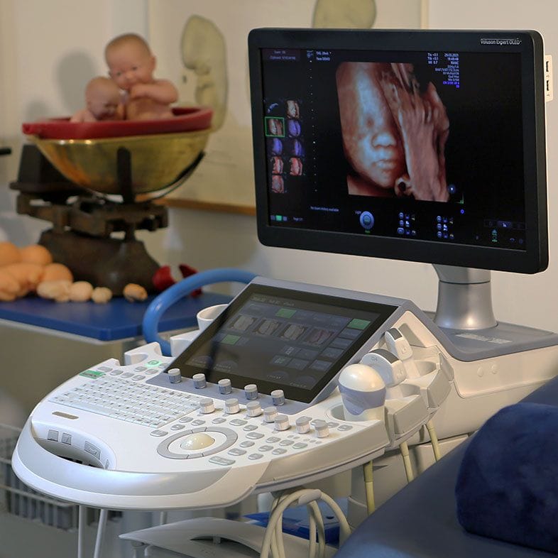
Ultrasound machine used to measure amniotic fluid volume during a pregnancy scan

Polyhydramnios in numbers

1–2% Polyhydramnios occurs in 1% to 2% of all pregnancies (StatPearls, 2024); UK maternity leaflets quote a wider 1–4% (Chelsea and Westminster NHS, 2024)
AFI ≥ 24 cm The amniotic fluid index adds the deepest pool in each of four quadrants; 24 cm or more is polyhydramnios (SMFM, 2018)
DVP ≥ 8 cm Or a single deepest vertical pocket of 8 cm or more — either measurement on its own is enough (SMFM, 2018)
65–70% About 65–70% of cases are mild, roughly 20% moderate and under 15% severe (StatPearls, 2024)
11% Of 672 pregnancies with polyhydramnios, 77 babies (11%) had an anomaly; ultrasound found nearly 80% of them (Dashe et al., 2002)
1% / 2% / 11% Anomaly risk left after a normal detailed scan: 1% with mild, 2% with moderate, 11% with severe polyhydramnios (Dashe et al., 2002)

How the fluid is measured, and what the numbers are

There are two accepted ways to measure amniotic fluid, and your report will use one. The amniotic fluid index (AFI) divides your bump into four quadrants, measures the deepest vertical pocket in each and adds the four figures. The deepest vertical pocket (DVP, or deepest pool) simply reports the largest pocket found anywhere.

Polyhydramnios is an AFI of 24 cm or more, or a DVP of 8 cm or more — either one alone is enough, and the Society for Maternal-Fetal Medicine uses exactly those thresholds. A normal deepest pocket sits between 2 cm and 8 cm; below 2 cm is the opposite problem, too little fluid.

Both numbers move — with your baby's position, with how recently they passed urine, and with who is holding the probe — so a reading a centimetre over the line is rarely acted on alone. A repeat scan a few weeks later is the normal response, and the second figure often comes back lower. Where a measurement does not fit, a second opinion scan re-measures.

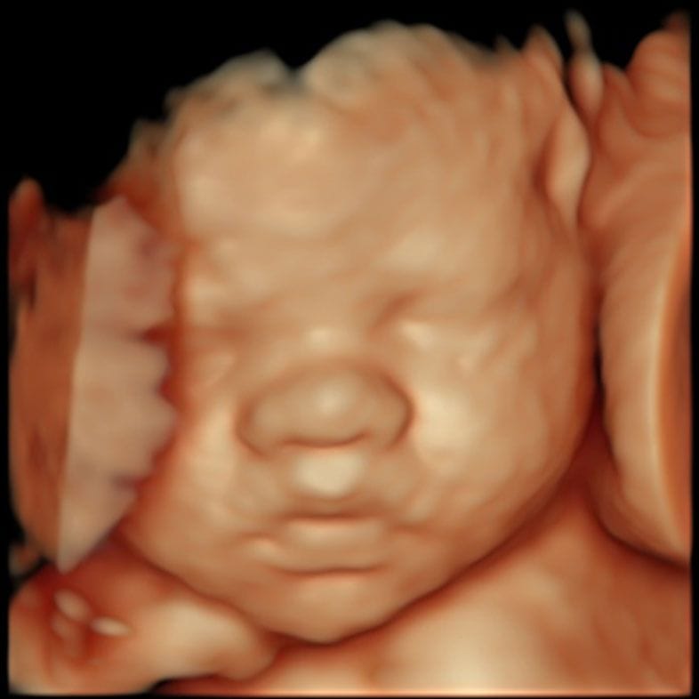
Third-trimester ultrasound image showing a baby and the pocket of fluid around them
Fluid volume varies between scans — the trend matters more than one figure

Mild, moderate and severe — what the bands mean

The band matters mainly because it changes how likely a cause is to be found. The last column is the one worth reading: the anomaly risk remaining after a detailed scan has looked and found nothing.

SeverityAmniotic fluid indexDeepest vertical pocketShare of casesAnomaly risk after a normal detailed scan
Mild24–29.9 cm8–11.9 cmAbout 65–70%About 1%
Moderate30–34.9 cm12–15.9 cmAbout 20%About 2%
Severe35 cm or more16 cm or moreUnder 15%About 11%

Bands and case-share: StatPearls (2024) and SMFM Consult Series #46 (2018). Residual anomaly risk: Dashe et al., 672 pregnancies with polyhydramnios, Obstetrics & Gynecology (2002). Dashe drew the mild band slightly higher, at an amniotic fluid index of 25.0–29.9 cm.

What is looked for — and how often it is found

Roughly in the order of how often each turns out to be the answer.

Nothing — idiopathic polyhydramnios

The commonest result, and the one most parents are eventually given. Estimates vary with severity: StatPearls puts idiopathic cases at 50–60% of the total; an Italian cohort found 177 of 356 (49.7%) unexplained; a 2025 South African series of 136 pregnancies found no cause in 81.2% of mild cases (65 of 80). Between half and four in five, then — a real finding with a good outlook, not a failed investigation.

Diabetes in pregnancy

The commonest treatable cause. Higher maternal glucose crosses to your baby, who makes more urine in response, and the fluid rises. In that 2025 series of 136 pregnancies, diabetes explained 15% of mild cases (12 of 80) and 16.7% of moderate cases (7 of 42). It is good news when it is the answer — controlling glucose usually settles the fluid. See gestational diabetes.

A problem with swallowing

If a baby cannot swallow and absorb fluid normally, it accumulates. The classic causes are oesophageal atresia — around 1 in 3,000 to 4,000 live births, with polyhydramnios present in 56.3% of cases — and duodenal atresia, a blockage past the stomach occurring in 1 in 5,000 to 10,000 live births and showing a 'double bubble' on ultrasound. Around 30% to 40% of babies with duodenal atresia have Down's syndrome. Both are repaired after birth.

Fetal anaemia

Uncommon, and looked for because it can be treated before birth. An anaemic baby's circulation speeds up, and blood flow in the middle cerebral artery can be measured with the same probe. It is checked particularly where fluid is collecting in the baby's own tissues.

Congenital infection

Some infections change how a baby handles fluid, so where polyhydramnios is unexplained a standard serology panel is checked or rechecked. Most results come back negative, and the screen is usually done for completeness — see TORCH screening and CMV testing.

Twins — twin-to-twin transfusion syndrome

In identical twins sharing one placenta, blood can flow unequally between them, leaving one twin with too much fluid and the other too little. It affects 8% to 10% of monochorionic twin pregnancies — which is why shared-placenta twins are scanned fortnightly from around 16 weeks.

What the workup involves

1

A detailed scan of your baby

The first and most useful step. Anatomy is worked through systematically — brain, face, chest, heart, stomach bubble, bowel, kidneys, limbs — alongside growth and the fluid measurement. In 672 such pregnancies, ultrasound identified nearly 80% of the babies who had an anomaly.

2

A glucose tolerance test

A two-hour oral glucose tolerance test through your maternity unit, offered even if you were screened earlier and passed, because tolerance drifts as pregnancy goes on. SMFM suggests rescreening may be considered where at least a month has passed since the last test.

3

Blood tests for infection, where the cause is unclear

Serology for syphilis, rubella, parvovirus B19, HIV, hepatitis, cytomegalovirus and toxoplasmosis. Not routine for every mild case — done where the polyhydramnios is unexplained, moderate or severe, or where something else shows.

4

A conversation about genetic testing, if the scan is not normal

In that same series, an extra or missing chromosome was found in 10% of babies with a structural anomaly on the scan, and in 1% of those without. Amniocentesis with a microarray is offered where the scan has found an anomaly. Where the polyhydramnios looks isolated, SMFM is clear that there are no data supporting amniocentesis for that reason alone — the test remains available to anyone who wants it, after a conversation about what it can and cannot tell you.

5

Repeat scans

Fluid is re-measured over time rather than judged once, with growth plotted alongside. Mild polyhydramnios with a normal scan and glucose test often needs nothing more than a recheck; moderate and severe cases are followed up in a fetal medicine unit.

The risks, said plainly

The reasons monitoring increases towards the end of pregnancy — the list your team is managing, not a list of things about to happen.

Waters breaking early, and preterm labour

A uterus stretched by extra fluid is more irritable, so membranes rupturing before term and preterm labour are both more likely. For context: in 109 pregnancies with mild idiopathic polyhydramnios against 2,550 controls, preterm birth was no more common (3.7% versus 3.1%).

Your baby not settling head-down

With more room to move, babies change position later and more freely, so an unstable or breech lie near term is more common. It is checked rather than assumed — presentation is confirmed by ultrasound before birth is planned. See our presentation scan.

Cord prolapse

If the waters break with a large volume of fluid while the head is still high, the cord can be swept down ahead of the baby. Uncommon, and an emergency when it happens — the reason you are told to ring the maternity unit the moment your waters break.

Bleeding after birth

An overstretched uterus does not always contract down briskly afterwards, so postpartum haemorrhage is more likely and your team will plan actively for the third stage. In the mild idiopathic group above it was no more common than in controls (9.2% versus 13.2%).

Stillbirth — the number, and the context

The figure people find at 2am, so here it is directly. In a Californian registry study of singleton pregnancies with no fetal anomaly, stillbirth occurred at 18.0 per 10,000 ongoing pregnancies with polyhydramnios at 37 weeks against 2.4 per 10,000 without, and 66.3 against 6.0 by 40 weeks — adjusted odds ratio 5.5 (95% CI 4.1–7.6). Several times higher, still well under 1 in 100, and why surveillance steps up towards term.

What mild polyhydramnios usually looks like

Caesarean birth is more likely; most other outcomes are not measurably different. For mild idiopathic polyhydramnios, SMFM does not recommend extra fetal surveillance for that reason alone, advises letting labour start spontaneously at term, and advises against induction before 39 weeks.

If your waters break, ring straight away

With extra fluid the gush can be sudden and large, and very occasionally the cord comes down with it. If your waters break, or you think they might have, phone your maternity unit immediately, and tell them you have polyhydramnios. The same applies to reduced movements at any hour, and to contractions before 37 weeks. None of these is likely; all are worth the call.

What a scan can see, and what it cannot

A scan measures fluid reliably and examines anatomy well, but it does not see everything. In the 672-pregnancy cohort ultrasound picked up nearly 80% of the babies who had an anomaly — good, not total. What most often slipped through were cardiac septal defects, cleft palate, imperforate anus and tracheo-oesophageal fistula. Oesophageal atresia is a particular blind spot: across 20 studies and 1,760 affected fetuses, prenatal ultrasound sensitivity was 31.7%.

A private scan cannot change how much fluid your baby makes, and no scan changes an outcome by itself. What it can do is measure the fluid and your baby on the day, look carefully at the anatomy polyhydramnios makes relevant, and give you a report your maternity team can act on — alongside NHS care, not instead of it. If the heart is the worry, see congenital heart defects. A growth scan or wellbeing scan also plots growth, so a baby measuring small as well is picked up — see fetal growth restriction.

Your questions answered

I've been told I have too much amniotic fluid — should I be worried?

Probably less than you currently are. It affects 1% to 2% of pregnancies, about 65% to 70% of cases are mild, and no cause is ever identified in around half of all cases — roughly four in five of the mild ones. The number that matters most: after a detailed scan has looked and found nothing, the chance of a major anomaly is about 1% with mild polyhydramnios and 2% with moderate (Dashe et al., 2002).

What causes polyhydramnios?

Most often nothing that can be found. Where there is a cause, diabetes in pregnancy and a problem with your baby swallowing are the commonest; less often fetal anaemia, a congenital infection, or twin-to-twin transfusion syndrome. In a 2025 series of 136 affected pregnancies, an anomaly was found in 2.86% of mild cases, rising to 21.4% of the 14 severe cases.

What is a normal amniotic fluid index?

Polyhydramnios starts at an amniotic fluid index of 24 cm or more, or a deepest vertical pocket of 8 cm or more — either alone is enough (SMFM, 2018). A normal deepest pocket is between 2 cm and 8 cm; below 2 cm is oligohydramnios. Because fluid shifts with your baby's position and with the person scanning, a figure just over the line is usually rechecked.

Does polyhydramnios mean something is wrong with my baby?

Usually not. In 672 pregnancies with polyhydramnios, 77 babies (11%) had an anomaly — meaning 89% did not — and ultrasound detected nearly 80% of those present (Dashe et al., 2002). It raises the question; it does not answer it.

Why do I need a glucose tolerance test when I already passed one?

Because glucose tolerance changes as pregnancy goes on, and polyhydramnios can be the first sign it has drifted. SMFM suggests rescreening may be considered where at least a month has passed since your last test. A positive result is one of the better outcomes of the workup — a treatable cause, and controlling glucose usually brings the fluid down.

Can polyhydramnios go away on its own?

Often it settles or fluctuates. Fluid volume is not fixed — it moves with your baby's position and with the person measuring, so a reading just over the threshold frequently comes back within range at the next scan. Where a cause has been found and treated, most often diabetes, the fluid commonly settles too.

Will I have to be induced early?

Not for mild idiopathic polyhydramnios on its own. SMFM's guidance is explicit: labour should be allowed to start spontaneously at term, and induction, if planned, should not happen before 39 weeks without another indication. Where it is severe, the plan is individual, and birth is advised in a unit with neonatal support on site.

Does polyhydramnios mean I will need a caesarean?

It makes one more likely, but most women still give birth vaginally. Comparing 109 pregnancies with mild idiopathic polyhydramnios against 2,550 controls, caesarean birth occurred in 46% versus 32%, emergency caesareans in 25.7% versus 17.7%. The usual reasons are the baby lying awkwardly and labour being less efficient in a stretched uterus.

Is there any treatment to reduce the fluid?

Rarely needed. Amnioreduction — draining 1.5 to 3 litres by needle under ultrasound guidance — is recommended only for severe polyhydramnios causing real discomfort or breathlessness, and more than one procedure is needed in 42% to 46% of cases because the fluid reaccumulates. Indomethacin reduces how much urine a baby makes, but SMFM advises against using it purely to lower fluid.

Does polyhydramnios increase the risk of stillbirth?

It raises it, and the absolute risk stays low. In a Californian registry study of singleton pregnancies with no fetal anomaly, the adjusted odds ratio was 5.5 (95% CI 4.1–7.6) — and even at 40 weeks the rate was under 1 in 100. That is why monitoring steps up towards term, and why pregnancies are not left far past the due date.

Will my baby be all right?

For most babies with polyhydramnios, yes — particularly where it is mild, the detailed scan is normal and the glucose test is negative, which describes most people reading this page. Where a cause is found, many are treatable: diabetes is managed, oesophageal and duodenal atresia are repaired after birth, fetal anaemia before it. No scan and no clinician can promise you an outcome; what monitoring buys is that decisions get made deliberately and in time. If you are frightened between appointments, ring your maternity unit — reduced movements warrant a call the same hour.

Contact

Speak to us about a growth scan

Send us an enquiry

A clinician will reply within one working day.

For urgent matters please call 020 3687 2939.

Sources & clinical references

The figures and clinical statements on this page are drawn from the sources below. Guidance evolves — always discuss your individual circumstances with a clinician.

  1. StatPearls / NCBI Bookshelf (Hwang, Jenkins, Mahdy)Polyhydramnios2024
  2. Society for Maternal-Fetal Medicine (SMFM)SMFM Consult Series #46: Evaluation and management of polyhydramnios2018
  3. Obstetrics & Gynecology (Dashe et al.), via PubMedHydramnios: anomaly prevalence and sonographic detection2002
  4. American Journal of Obstetrics and Gynecology, via PubMedThe risk of fetal death in nonanomalous pregnancies affected by polyhydramnios2015
  5. PLOS ONE (Siveregi, Horak, Stewart)Causes, management and outcomes of polyhydramnios at a secondary level hospital in Cape Town, South Africa2025
  6. Children (Basel) (Pasquini et al.), via PubMed CentralObstetric and Neonatal Outcomes in Mild Idiopathic Polyhydramnios2022
  7. Acta Obstetricia et Gynecologica Scandinavica (Pardy et al.), via PubMedPrenatal detection of esophageal atresia: A systematic review and meta-analysis2019
  8. StatPearls / NCBI Bookshelf (Sigmon, Eovaldi, Cohen)Duodenal Atresia and Stenosis2023
  9. Neonatal Network NHS ScotlandOesophageal atresia — information for patients and families2024
  10. Chelsea and Westminster Hospital NHS Foundation TrustPolyhydramnios — patient information2024