
Ventriculomegaly

If a scan has just shown ventriculomegaly, start here: the fluid-filled spaces inside your baby's brain measure a little wider than the usual cut-off, and in the mild isolated form that is usually all it means. Around 90% of babies with a mild measurement of 10–12mm develop normally; about 10% have neurodevelopmental delay (NHS Genomics Education Programme) — and where the finding is genuinely isolated, UK guidance says the risk may not be dramatically higher than the background population risk (NHS Greater Glasgow and Clyde). The work now is establishing whether the finding really is isolated, and that is what a detailed anomaly scan is for.

What ventriculomegaly actually means

Ventriculomegaly is a measurement, not a diagnosis. The ventricles are the fluid-filled spaces inside the brain — every brain has them, and they are meant to contain fluid. When one measures 10mm or more at the point where it is routinely measured, that word goes in your notes. Nothing more has been said about your baby than that.

It is not a hole in the brain, and on its own it is not brain damage. Cambridge University Hospitals tells parents the fluid normally measures less than 10mm at 20 weeks, and that the appearance is seen in about 1 in every 250 pregnancies at that scan. Newcastle upon Tyne Hospitals is equally direct: most babies measuring 10mm or more are completely normal, the wider space simply reflecting natural variation in how a brain develops.

It matters when the wider ventricles are a signpost to something else — an infection passed to your baby, a chromosomal difference, another structural difference in the brain, or a bleed. That is why the answer is a careful look at everything else, not a repeat of the same measurement. A detailed anomaly scan, or a second opinion scan if you want it re-measured, exists to answer that question.

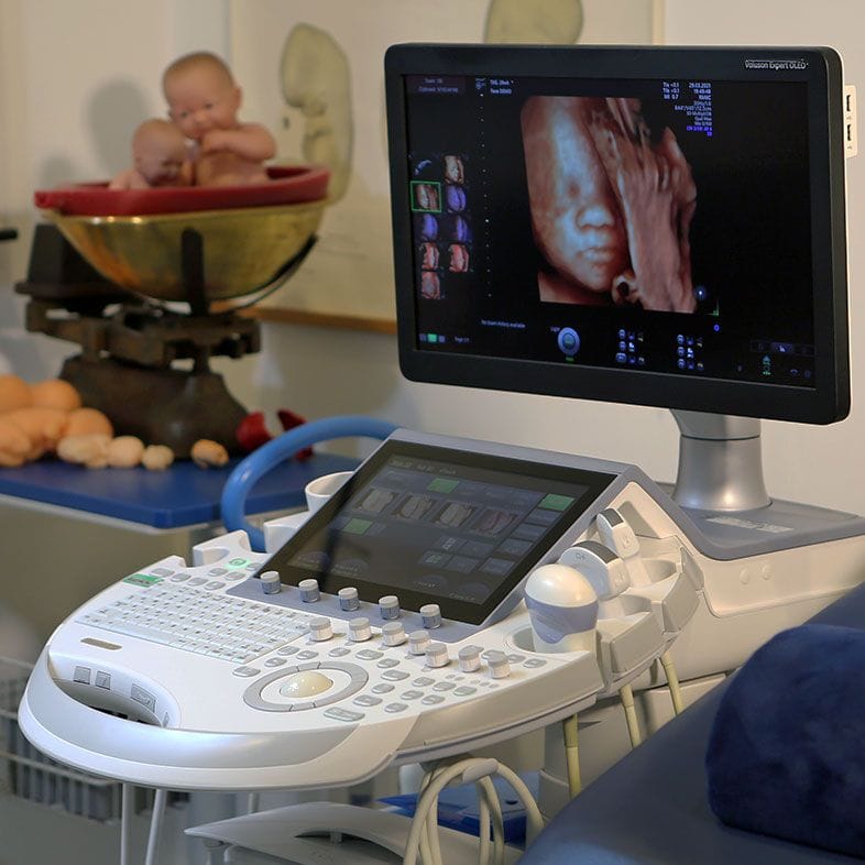
High-resolution ultrasound equipment used for detailed fetal brain assessment
Millimetres decide the label, so technique matters

Ventriculomegaly in numbers

10mm Ventriculomegaly is an atrial measurement of 10mm or more in the lateral ventricle (NHS Greater Glasgow and Clyde)
1–2 in 1,000 Seen in 1–2 per 1,000 pregnancies, though some UK units quote nearer 1 in 250 at the 20-week scan (NHS Genomics Education Programme; Cambridge University Hospitals)
About 90% About 90% of babies with mild ventriculomegaly of 10–12mm develop normally; about 10% have neurodevelopmental delay (NHS Genomics Education Programme)
50–60% One ventricle only is affected in 50–60% of cases and both in 40–50%, so both sides are always measured (NHS Greater Glasgow and Clyde)
0–5% An abnormal karyotype is found in 0–5% of apparently isolated mild cases, with a further 10–15% having changes on microarray (NHS Greater Glasgow and Clyde)
5–6% Fetal MRI finds clinically relevant extra brain findings ultrasound missed in about 5–6% of cases in recent series, against a published range of 1–14% (NHS Greater Glasgow and Clyde)

The measurement, and where the 10mm line comes from

The figure everyone quotes is the atrial width — the back part of the lateral ventricle. It is taken on a standard axial view of the head, at the level of the frontal horns and the cavum septum pellucidum. The callipers go on the inner margins of the ventricle walls, at the parieto-occipital groove and the glomus of the choroid plexus, perpendicular to the long axis of the ventricle (NHS Greater Glasgow and Clyde).

Under 10mm is normal, and that threshold holds steady through the second and early third trimesters — which is why a figure taken today can be compared with one taken six weeks from now. It also means a millimetre or two between scans can reflect who held the probe. That is one reason a borderline measurement is often simply repeated by someone with fetal neurosonography experience before anything else is set in motion.

Both ventricles are measured, deliberately: the one further from the probe is easier to see, so a one-sided finding has to be checked rather than assumed. Between 50 and 60% of cases involve one ventricle only, and 40 to 50% involve both (NHS Greater Glasgow and Clyde). A unilateral finding carries a slightly lower reported risk than a bilateral one.

Sonographer performing a detailed fetal anomaly scan of the baby's head and brain

Mild, moderate and severe

The bands describe how far past the cut-off the measurement sits, not a prediction for your baby. The figures below are for ventriculomegaly found on its own; where it is not isolated, or where it progresses, the picture changes and is set out by your fetal medicine team case by case.

BandAtrial widthWhat the outcome data show
Mild10–12mmAbout 90% develop normally, about 10% have neurodevelopmental delay (NHS Genomics Education Programme). NHS Greater Glasgow and Clyde puts the risk at about 6% when one ventricle is affected and 8–12% when both are, adding that this may not be dramatically higher than the background population risk.
Moderate13–15mmAlso put at about 90% developing normally by the NHS Genomics Education Programme. A 2024 review gives a wider, more cautious range of 75–93% favourable outcomes (Children, Zamłyński et al.).
SevereOver 15mmAbout 50% have neurodevelopmental delay and/or severe disability (NHS Genomics Education Programme). In a 2024 meta-analysis of isolated severe cases, 73 of 125 surviving infants (58.4%) had adverse neurodevelopment and 74 of 304 (24.3%) died in utero (Translational Pediatrics, Ali et al.).

Definitions differ slightly. ISUOG's patient information treats 10–15mm as ventriculomegaly, calls up to 12mm mild, and uses hydrocephaly above 15mm; UK unit guidance more often uses 10–12mm, 13–15mm and over 15mm. The 10mm threshold itself is not in dispute.

Most families are told a number in the mild band. Referral series look worse than reality because they collect the hard cases: of 229 fetuses referred to two fetal medicine centres, 47.6% were mild, 26.2% moderate and 26.2% severe (Prenatal Diagnosis, Moens et al., 2025).

Why ‘isolated’ is the word doing all the work

Almost every reassuring figure on this page has the word isolated in front of it. Isolated means that after a thorough look, the wider ventricles are the only finding: nothing else in the brain, nothing structural elsewhere, no evidence of infection, no chromosomal cause. That is the group with the good outcomes — and it is a different group from ‘nothing else has been noticed yet’.

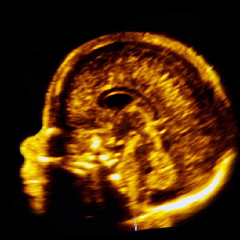
Establishing isolation is what a detailed scan is for, and it is a different examination from measuring the ventricle again. It is a deliberate survey of the rest of the brain — corpus callosum, cerebellum and posterior fossa, cortex and its folding pattern, septum pellucidum — then the spine, heart, kidneys and growth, because ventriculomegaly is sometimes the visible end of something systemic. Transvaginal ultrasound is often used when the baby is head-down, because the probe gets closer to the brain.

UK guidance states the honest limit rather than hiding it: most published statistics rest on whether ventriculomegaly is apparently isolated, and true isolation can only be confirmed after birth (NHS Greater Glasgow and Clyde). A scan reduces uncertainty; it does not abolish it. Our page on common fetal anomalies sets out what else a full survey covers.

What a UK fetal medicine unit will do next

Five things follow a measurement of 10mm or more, and they all serve the same question — is this isolated? Every one of them is a choice you can decline.

Detailed neurosonography

A dedicated examination of the fetal brain by someone who does it regularly, often transabdominal and transvaginal combined depending on how your baby is lying. The midline structures, posterior fossa, cortex and the shape of the ventricles are all assessed, not just the atrial width, and the rest of the body is surveyed at the same time. This is the step that decides whether the word isolated can be used at all.

Infection screening

A blood test on you, not on your baby. NHS Greater Glasgow and Clyde advises offering tests for cytomegalovirus (CMV), toxoplasmosis and parvovirus regardless of known exposure or symptoms, because these infections in pregnancy are usually silent. That guidance associates congenital infection with mild ventriculomegaly in around 8% of cases; a 2024 review puts it lower, at about 1.4%. Either way, worth excluding early.

Karyotype and microarray

Invasive testing, usually amniocentesis, is offered so the chromosomes can be examined directly and a microarray run. In apparently isolated mild ventriculomegaly, 0–5% of babies have an abnormal karyotype — most commonly Down's syndrome — and a further 10–15% have a change found on microarray (NHS Greater Glasgow and Clyde). Amniocentesis carries a miscarriage risk quoted by Cambridge University Hospitals at about 0.5%, roughly 1 in 200. Declining is a legitimate choice.

Fetal MRI

MRI uses no radiation and is arranged through a fetal medicine department, to look for brain findings ultrasound cannot resolve. The chance of it finding a clinically relevant additional abnormality ranges across the literature from 1% to 14%, with recent studies at 5–6% (NHS Greater Glasgow and Clyde) — the figure falls as the quality of the initial neurosonography rises. The commonest additional finding is agenesis of the corpus callosum.

Repeat scans

There is no evidence base for the ideal interval; UK guidance suggests a pragmatic four-weekly assessment. What is watched is the direction of travel, not the absolute number, because progression is an important prognostic indicator. Most measurements do not get worse. If yours does, that changes the conversation — which is why the follow-up exists, not a reason to expect it.

What a scan can and cannot tell you

An ultrasound can measure the ventricles precisely, survey the rest of the brain and body, and tell you whether anything else is visible today. It cannot tell you how your child will develop, and no scan — NHS or private, ultrasound or MRI — changes what is happening inside your baby's head. Imaging buys information, sometimes sooner. It does not buy a different outcome.

When to call your maternity unit instead of booking a scan

Ventriculomegaly itself does not make you feel unwell, so anything you do feel needs assessing on its own terms. Reduced or altered fetal movements, a fever or flu-like illness, a rash after contact with someone unwell, bleeding, abdominal pain or any fluid loss all warrant a call to your maternity unit or triage the same day — they are open around the clock and would far rather hear from you. A private scan is for a second opinion or a longer look. It is never a substitute for urgent obstetric assessment, and you should not sit on a symptom while you wait for one.

Ventriculomegaly is not the same as hydrocephalus

These two words get used interchangeably online and they are not interchangeable. Ventriculomegaly is a measurement: a ventricle is wider than 10mm. Hydrocephalus is a mechanism: fluid is building up and pressing on the brain, which the NHS describes as capable of damaging it. Most babies with ventriculomegaly do not have hydrocephalus, and a mild isolated measurement of 10 or 11mm is a long way from it.

The words converge only at the severe end. ISUOG's patient information uses hydrocephaly above 15mm, where the ventricles are not merely wide but expanding, often because something is obstructing the flow of fluid. Babies born with hydrocephalus are usually treated with a shunt — a thin tube placed surgically to drain the excess fluid — or sometimes an endoscopic third ventriculostomy (NHS). If that is the conversation you are actually having, our hydrocephalus page covers it in more detail.

This is worth spelling out because a 2am search for ‘fluid on baby’s brain’ returns hydrocephalus material within seconds, written for a different situation from yours.

What happens between now and birth

Most measurements do not get worse

In 324 cases of mild ventriculomegaly found among 40,192 singleton pregnancies at one Beijing centre, the measurement regressed in 57–58% of babies, stayed stable in 28–36% and progressed in 6–15%, depending on whether it was picked up early or late in pregnancy (European Journal of Medical Research, Wang et al., 2024). NHS Greater Glasgow and Clyde quotes progression at around 5%. Referral series report higher rates because they see the harder cases.

When it does progress

Progression matters more than the starting number, which is why it is the thing being watched at roughly four-weekly scans. Across 229 referred fetuses at two centres, among those followed up the ventriculomegaly resolved altogether in 37.3% (57 of 153) and progressed in 29.4% (45 of 153); progression was commonest in babies whose measurement was already severe, where 51.9% progressed (Prenatal Diagnosis, Moens et al., 2025). A rising measurement is a reason to look again for a cause, not a verdict.

Birth, and afterwards

For isolated mild ventriculomegaly, birth is planned on ordinary obstetric grounds — the finding itself does not mean induction, early delivery or a caesarean. The neonatal team is told in advance, cord bloods can be taken with your consent for chromosomal and infection testing if that was not done in pregnancy, and paediatric follow-up is arranged before you leave hospital (NHS Greater Glasgow and Clyde). Postnatal imaging occasionally finds something not visible before birth.

Where a private scan fits

A second opinion scan can re-measure the ventricles, survey the brain and the rest of your baby in detail, and give you a written report to hand to your NHS team. It does not replace your NHS fetal medicine pathway and it cannot change the measurement. What it can do is shorten the wait between being told something and understanding it. Dr Fred Ushakov sees these referrals directly.

Your questions answered

They said there is fluid on my baby's brain — is my baby going to be OK?

The phrase frightens people more than the finding warrants. There is meant to be fluid in those spaces; the scan has found them slightly wider than the 10mm cut-off. If the measurement is mild and nothing else is found, about 90% of babies develop normally and about 10% have some neurodevelopmental delay (NHS Genomics Education Programme).

Where the finding is genuinely isolated, UK guidance puts the risk nearer 6% if one ventricle is affected and 8–12% if both are, and says that may not be dramatically higher than the background population risk (NHS Greater Glasgow and Clyde). Nobody can promise you an outcome, but the honest starting position is a reassuring one.

What does 10mm on a brain scan actually mean?

It is the width of the atrium — the back part of the lateral ventricle — on a standard view of the head. Under 10mm is normal throughout the second and early third trimesters; at 10mm or above, the word ventriculomegaly is used. A millimetre either side of that line can come down to where the callipers were placed, which is why a borderline number is often simply re-measured.

Is mild ventriculomegaly serious?

Usually not, when it is isolated. UK guidance puts the risk of neurodevelopmental delay at about 6% for isolated mild or moderate ventriculomegaly affecting one ventricle, and 8–12% when both are affected, adding that this may not be dramatically higher than the background population risk (NHS Greater Glasgow and Clyde).

ISUOG says the risk of neurological impairment is no higher than 10% when the finding is mild and isolated.

Those figures stop applying the moment it is not isolated — if anything else is found in the brain or body, an infection or chromosomal cause is identified, or the measurement progresses, the outlook is different and your fetal medicine team will give you numbers for your situation rather than these.

Can ventriculomegaly go away on its own?

Often, yes. Among 324 babies with mild ventriculomegaly found in 40,192 singleton pregnancies at one Beijing centre, the measurement regressed in 57–58% and stayed stable in a further 28–36% (European Journal of Medical Research, Wang et al., 2024). In a referral series of 229 fetuses including moderate and severe cases, it resolved altogether in 37.3% of those followed up (57 of 153) (Prenatal Diagnosis, Moens et al., 2025). A realistic hope, not a promise anyone can make at the first scan.

Is ventriculomegaly the same as hydrocephalus?

No. Ventriculomegaly is a measurement above 10mm. Hydrocephalus means fluid is accumulating under pressure and pressing on the brain. Most babies with ventriculomegaly do not have hydrocephalus. The terms only start to overlap above 15mm, which ISUOG calls hydrocephaly. If your number is in the mild band, the hydrocephalus pages you have probably already found describe a different situation.

Does ventriculomegaly mean my baby has Down's syndrome?

Almost always no, but it raises the question enough that testing is offered. In apparently isolated mild ventriculomegaly, 0–5% of babies have an abnormal karyotype, Down's syndrome being the commonest, and a further 10–15% have a change found on microarray (NHS Greater Glasgow and Clyde). A low-risk NIPT result already makes the common trisomies much less likely, but it does not look for the microarray findings.

Do I have to have an amniocentesis?

No. It is offered, not required, and plenty of parents decline — particularly when the measurement is mild, the detailed scan is otherwise normal and screening has been reassuring. What amniocentesis adds is a definitive chromosomal answer including microarray. What it costs is a procedure-related miscarriage risk of about 0.5%, roughly 1 in 200 (Cambridge University Hospitals). It is reasonable to take a few days over that decision.

Why are they testing my blood for CMV and toxoplasmosis?

Because both can cause ventriculomegaly, and both can pass to a baby without the mother ever feeling unwell. UK guidance advises offering CMV, toxoplasmosis and parvovirus testing regardless of known exposure or symptoms (NHS Greater Glasgow and Clyde). That guidance links congenital infection to around 8% of mild cases; a 2024 review puts it nearer 1.4%. A negative result removes one of the more consequential explanations.

Do I need a fetal MRI?

Not everyone does. MRI is an adjunct to ultrasound, not a replacement, and its value depends on how good the initial brain scan was. The chance it finds an important brain abnormality ultrasound missed ranges from 1% to 14% across the literature, with recent studies at 5–6% (NHS Greater Glasgow and Clyde) — the better the neurosonography, the lower that number. Ask your team what they expect it would change.

Will my baby need a shunt?

Shunts treat hydrocephalus, not mild ventriculomegaly, and the overwhelming majority of babies with a mild isolated measurement never need neurosurgical treatment. Where hydrocephalus does develop, a shunt drains the excess fluid and an endoscopic third ventriculostomy is sometimes used instead (NHS). This is a question for the severe end of the range, not for a 10 or 11mm finding.

Did I do something to cause this?

No. Nothing you ate, lifted, drank before you knew, or worried about made your baby's ventricles measure 11mm instead of 9mm. The listed causes are normal anatomical variation, congenital infection, chromosomal and genetic conditions, structural brain differences and bleeding within the brain (NHS Greater Glasgow and Clyde) — and by a distance the commonest is normal variation. Parents ask this at almost every one of these appointments, so ask it out loud.

What should I ask at my next appointment?

What exactly did each ventricle measure, and on which side? Has anything else at all been seen in the brain or body? Which of infection screening, invasive testing and MRI are you recommending for me, and what would each change? When am I scanned again? Write the millimetres down — the actual numbers make every later conversation, including a second opinion scan, far more useful.

Contact

Speak to us about a detailed brain scan

Send us an enquiry

A clinician will reply within one working day.

For urgent matters please call 020 3687 2939.

Sources & clinical references

The figures and clinical statements on this page are drawn from the sources below. Guidance evolves — always discuss your individual circumstances with a clinician.

  1. NHS Genomics Education Programme (GeNotes)Ventriculomegaly — Knowledge Hub2024
  2. NHS Greater Glasgow and Clyde (Right Decisions clinical guidelines)Mild Ventriculomegaly on Antenatal Ultrasound (916)2024
  3. International Society of Ultrasound in Obstetrics and Gynecology (ISUOG)Ventriculomegaly — patient information series2024
  4. Newcastle upon Tyne Hospitals NHS Foundation TrustMy baby has ventriculomegaly on ultrasound scan2023
  5. Cambridge University Hospitals NHS Foundation TrustVentriculomegaly — patient information2024
  6. Translational Pediatrics (Ali, Gurung, Nanda, Bakalis, Sankaran, Arichi, Nicolaides, Shangaris), via PubMed CentralPerinatal and neurodevelopmental outcomes of fetal isolated ventriculomegaly: a systematic review and meta-analysis2024
  7. Children (Zamłyński, Zhemela, Olejek), via PubMed CentralIsolated Fetal Ventriculomegaly: Diagnosis and Treatment in the Prenatal Period2024
  8. European Journal of Medical Research (Wang et al.), via PubMed CentralFollow-up outcome analysis of 324 cases of early-onset and late-onset mild fetal ventriculomegaly: a retrospective cohort study2024
  9. Prenatal Diagnosis (Moens et al.), via PubMed CentralClinical Outcome and Risk Factors for Progression of Prenatally Diagnosed Fetal Ventriculomegaly: A Retrospective Multicenter Study2025
  10. NHSHydrocephalus2023