Choroid Plexus Cyst

A choroid plexus cyst is a small bubble of fluid inside the choroid plexus — the tissue in your baby’s brain that makes the fluid the brain and spinal cord float in. It is not a brain abnormality, it is not in the thinking part of the brain, and in babies with normal chromosomes it has not been shown to affect brain development or intelligence. Cysts like this are seen in about 1 to 2 of every 100 babies, and most have gone by 26 to 28 weeks.

What a choroid plexus cyst actually is

If a sonographer has just told you there is a cyst on your baby’s brain, here is the part that matters most: a choroid plexus cyst is not a hole, a growth or damage, and it is not in the part of the brain that does the thinking. The choroid plexus is spongy tissue inside the fluid-filled spaces in the middle of the brain, and its job is to make cerebrospinal fluid — the clear fluid that cushions the brain and spinal cord (ISUOG). A cyst is a tiny pocket of that fluid trapped inside the tissue that makes it.

Because the choroid plexus is a fluid factory rather than a thinking structure, a cyst in it does not press on anything that matters and has not been shown to interfere with how the brain forms. The Society for Maternal-Fetal Medicine describes an isolated cyst — one found with nothing else unusual on the scan — in a pregnancy with a negative chromosome screening result as a normal variant of no clinical importance, needing no follow-up scan and no check after birth.

Cysts of this kind are found in about 1 to 2 of every 100 babies (ISUOG), which makes them one of the more common things a sonographer sees when looking at the fetal brain at the 20-week anomaly scan. They may be on one side or on both. The question worth asking is not how big the cyst is, but whether it is isolated — that is what current guidance turns on, and for the great majority of parents the answer is yes.

Detailed fetal ultrasound assessment of the baby’s brain and anatomy

Choroid plexus cysts in numbers

Every figure below is drawn from the sources listed at the foot of this page.

1–2 in 100 Choroid plexus cysts are seen in about 1 to 2 of every 100 babies, most often on second-trimester scans (ISUOG)
26–28 weeks The NHS Perinatal Institute describes cysts as almost always gone by 26 weeks; ISUOG says most resolve by 28 weeks
2.1% Of all babies found to have a cyst, 2.1% have a chromosome difference — and most of those have other abnormalities visible on the same scan (ISUOG)
7 in 1,346 Across 246,545 second-trimester scans, 7 of 1,346 babies with an isolated cyst had trisomy 18, about 1 in 190 (Yoder et al., 1999)
87% Of 69 confirmed trisomy 18 pregnancies, 87% had two or more abnormalities at the 18 to 21 week scan, so a cyst was rarely the only sign (Lai et al., 2010)
1 in 1,152 Edwards’ syndrome affected about 1 in 1,152 total births in England in 2022, and about 1 in 11,315 live births (NHS England, NCARDRS)
No difference A 2023 review of eight studies found no difference in development or physical health in childhood after an isolated cyst (Singal et al., 2023)

Why a cyst appears, and why it goes

Choroid plexus cysts form as part of ordinary development. In the first half of pregnancy the tissue grows quickly and its surface is folded and loose, so small pockets of cerebrospinal fluid get trapped between the folds and show on ultrasound as neat round dark circles. Nothing has gone wrong to produce them.

They are commonest at around 16 weeks and get harder to see as pregnancy goes on. The Perinatal Institute, an NHS body, describes them as almost always disappearing by 26 weeks; ISUOG’s patient information says most resolve by 28 weeks. The two differ slightly, so 26 to 28 weeks is the honest range, and by birth there is usually nothing left to find.

Because they go on their own, the cyst needs no draining, treating or monitoring. Where it is the only finding and chromosome screening is negative, the Society for Maternal-Fetal Medicine says there is no indication for a repeat scan and none for examining the baby afterwards.

The longer view is reassuring too. A 2023 systematic review pooled eight studies of children who had had an isolated choroid plexus cyst seen before birth; across the three cohort studies there was no difference in developmental outcomes or physical health (Singal et al., 2023). The authors noted the studies were small and that attention, learning and epilepsy deserve larger research — so this is reassuring evidence rather than a guarantee — but nothing published so far points to harm.

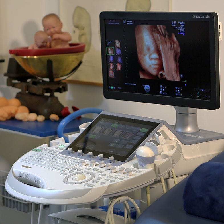
Ultrasound equipment used for detailed assessment of the fetal brain
Cysts are easiest to see around 16 weeks and usually fade as pregnancy goes on

Isolated cyst, or a cyst alongside something else

These are not the same finding, and the difference between them is the whole story. Ask your team which one applies to you.

The usual picture

An isolated choroid plexus cyst

“Isolated” means the sonographer has checked the rest of your baby — brain, face and profile, heart and its outflow tracts, hands and fingers, abdominal wall, feet and growth — and everything else is normal. This is the situation for the overwhelming majority of cysts.

The Society for Maternal-Fetal Medicine treats an isolated cyst with a negative screening result as a normal variant of no clinical importance, needing no follow-up scan and no newborn check. If you have not yet had screening, the recommended next step is counselling about your chance of Edwards’ syndrome and the offer of NIPT or a quadruple blood test — not an invasive test.

Needs specialist review

A cyst with another finding

If the cyst is seen with something else — a heart abnormality, clenched hands with overlapping fingers, a small chin, an exomphalos, clubbed or rocker-bottom feet, a small head, or a baby measuring behind — this is no longer a soft marker standing on its own. That combination is what genuinely raises concern about Edwards’ syndrome, and it is a reason to be seen promptly by a fetal medicine specialist and offered diagnostic testing such as amniocentesis, which examines the baby’s own chromosomes. The cyst is the least important item on that list.

The Edwards’ syndrome question, answered honestly

The only reason choroid plexus cysts get mentioned at all is a reported link with Edwards’ syndrome, or trisomy 18. It is worth setting the numbers out plainly, because a vague answer is more frightening than a specific one.

Edwards’ syndrome is uncommon. In England in 2022 the total birth prevalence was 8.7 per 10,000 births — about 1 in 1,152 — and because many affected pregnancies do not continue to term, the live birth prevalence was 0.9 per 10,000, about 1 in 11,315 (NHS England, NCARDRS).

Babies with trisomy 18 do have choroid plexus cysts about a third of the time (ISUOG), which is where the association comes from — but that statistic runs in the direction that does not help you. Turned to face the question you are actually asking: of all babies found to have a cyst, 2.

1% have a chromosome difference, and most of those babies have other abnormalities visible on the same scan (ISUOG). For an isolated cyst, a meta-analysis of 13 prospective studies covering 246,545 second-trimester scans found 7 cases of trisomy 18 among 1,346 babies — about 1 in 190 — with a summary likelihood ratio of 13.8 (95% CI 7.72 to 25.14) (Yoder et al., 1999).

ISUOG puts the residual chance nearer 1 in 300. The two estimates differ, so take them as a range; on either, well over 99% of babies with an isolated cyst do not have trisomy 18.

There is no comparable link with Down’s syndrome. In the same analysis the likelihood ratio for trisomy 21 was 1.87, with a confidence interval of 0.78 to 4.46 — crossing 1, which means the finding did not shift the odds in any statistically convincing way.

The second reason an isolated cyst is reassuring is that trisomy 18 rarely travels alone. In a series of 69 confirmed cases, 87% of babies had two or more abnormalities visible at the 18 to 21 week anomaly scan, and the reported detection rate of that scan was 100% (Lai et al.

, 2010); the Perinatal Institute describes additional ultrasound features in around 80% of trisomy 18 pregnancies. A cyst on its own, in a baby whose heart, hands, profile, abdominal wall and growth all look normal, is a very different finding from a cyst in that company. No scan can exclude every condition, but a careful anatomy survey is where the useful information lives.

This is why current guidance no longer treats an isolated cyst as a reason for an invasive test. Where screening with cell-free DNA or a quadruple test has already come back negative, the Society for Maternal-Fetal Medicine recommends no further evaluation for chromosome conditions.

Where no screening has been done, it recommends counselling to estimate your individual chance of trisomy 18 and a discussion of NIPT or a quadruple screen. A meta-analysis of cell-free DNA accuracy found a detection rate of 97.4% (95% CI 95.8 to 98.4) for trisomy 18 with a specificity of 99.9% (Taylor-Phillips et al.

, 2016) — but NIPT is a screening test, and a high-chance result needs amniocentesis to confirm it.

What to do next

Ask whether the cyst is isolated

The most useful question you can ask, and worth asking before you leave or on a follow-up call. “Isolated” means the rest of the anatomy — brain, face, heart and its outflow tracts, hands and fingers, abdominal wall, feet and growth — has been checked and looks normal. If some views could not be obtained, that is common and usually means another look rather than bad news.

Check the screening you have had

If you have already had NIPT or the combined or quadruple test with a low-chance result, guidance says an isolated cyst adds nothing that needs acting on. If you have not, genetic counselling can help you decide whether you want a screening test at all, and what a result would and would not tell you.

Have the anatomy looked at, not the cyst

Where trisomy 18 is the concern, the value of another scan lies in everything other than the cyst: the heart and its outflow tracts, the hands and fingers, the profile and chin, the abdominal wall, the feet and growth. A second opinion scan is a way to have that anatomy reviewed independently. It cannot promise a particular result, and no scan rules out every condition.

Screening is not diagnosis

NIPT is a screening test run on a blood sample from you: for trisomy 18, a detection rate of 97.4% with a specificity of 99.9% (Taylor-Phillips et al., 2016). It cannot give a diagnosis. Only amniocentesis or CVS analyses your baby’s own chromosomes, and each carries a small procedure-related risk that should be explained to you fully first.

Repeat scans for the cyst itself

Where the cyst is isolated and screening is negative, the Society for Maternal-Fetal Medicine says no follow-up ultrasound imaging and no postnatal evaluation are indicated. Some units still offer a later look to confirm growth is tracking normally. If you would simply like to see your baby again, a growth scan is available — your choice, not a clinical requirement.

Support while you wait

Waiting is the hardest part, and it is normal for this to sit with you for days. Antenatal Results and Choices (ARC) is a UK charity with a helpline for exactly this situation, and your midwife or screening team can arrange a conversation with a fetal medicine specialist. To talk it through here, an appointment with Dr Fred Ushakov or a genetic counsellor can be arranged.

Your questions answered

They’ve found a cyst on my baby’s brain — is it serious?

In almost all cases, no. A choroid plexus cyst is a small pocket of fluid inside the tissue that makes the brain’s fluid. It is not a hole in the brain, not a tumour and not damage, and it is not in the part of the brain used for thinking, memory or movement. Cysts of this kind are found in about 1 to 2 of every 100 babies (ISUOG).

The Society for Maternal-Fetal Medicine describes an isolated cyst — one found with nothing else unusual on the scan — with a negative chromosome screening result as a normal variant of no clinical importance. The one thing worth clarifying with your team is whether anything else was seen, because that is what changes the answer.

Will a choroid plexus cyst affect my baby’s development or intelligence?

There is no evidence that it does. A 2023 systematic review pooled eight studies of children who had had an isolated choroid plexus cyst detected before birth, and across the three cohort studies there was no difference in developmental outcomes or physical health (Singal et al., 2023). ISUOG likewise reports no differences in brain function, motor function or behaviour in children with normal chromosomes. The authors noted the studies were small, so the fair way to put it is that nothing published so far points to harm.

What causes a choroid plexus cyst — did I do something wrong?

No. Choroid plexus cysts arise as part of normal development: the choroid plexus grows quickly in the first half of pregnancy and its folded surface traps small pockets of cerebrospinal fluid. They are not known to be caused by anything a parent did or did not do in pregnancy, there is nothing you could have taken or avoided that would have prevented one, and they are not inherited in any recognised way.

How common are choroid plexus cysts?

ISUOG puts the figure at 1 to 2% of babies, roughly 1 in every 50 to 100. The Perinatal Institute reports about 2% of fetuses showing the finding at 16 weeks, which is when cysts are easiest to see. That commonness is part of the reassurance: if cysts were a reliable sign of a serious problem, they would not appear in one or two babies out of every hundred scanned.

Will the cyst go away on its own?

Almost always. The Perinatal Institute describes them as almost always gone by 26 weeks and ISUOG says most resolve by 28 weeks, so 26 to 28 weeks is the honest range, and by birth there is usually nothing left to see. They need no draining or treatment, and where the cyst is isolated with negative screening there is no indication for a follow-up scan just to watch it disappear.

Does a choroid plexus cyst mean my baby has Edwards’ syndrome?

Almost certainly not. Babies with trisomy 18 do have choroid plexus cysts about a third of the time, which is why the association exists — but very few babies with a cyst have trisomy 18. ISUOG reports that 2.1% of all babies found to have a cyst have a chromosome difference, and that most of those babies have other abnormalities visible on the same scan.

For an isolated cyst the numbers are smaller again: 7 cases of trisomy 18 among 1,346 babies in a meta-analysis of 246,545 second-trimester scans, about 1 in 190 (Yoder et al., 1999), and nearer 1 in 300 on ISUOG’s estimate. On either figure, well over 99% of babies with an isolated cyst do not have Edwards’ syndrome.

Is a choroid plexus cyst linked to Down’s syndrome?

Not in any convincing way. In the meta-analysis of 246,545 second-trimester scans, the likelihood ratio for trisomy 21 in babies with an isolated choroid plexus cyst was 1.87, with a confidence interval of 0.78 to 4.46 (Yoder et al., 1999). Because that interval crosses 1, the finding did not meaningfully change the chance of Down’s syndrome. If you are weighing up screening, that decision stands on its own merits rather than on the cyst.

My NIPT came back low chance — do I still need to worry about the cyst?

This is the clearest situation in the guidance. For an isolated choroid plexus cyst with a negative cell-free DNA or serum screening result, the Society for Maternal-Fetal Medicine recommends no further evaluation for chromosome conditions, because the finding is a normal variant of no clinical importance — with no indication for a follow-up scan or any check after birth.

For context, a meta-analysis found cell-free DNA testing detected 97.4% of trisomy 18 cases with a specificity of 99.9% (Taylor-Phillips et al., 2016). NIPT is not a diagnostic test, but a low-chance result alongside an otherwise normal anatomy scan is a genuinely reassuring combination.

Do I need an amniocentesis?

For an isolated cyst, current guidance does not recommend going straight to an invasive test. The Society for Maternal-Fetal Medicine recommends counselling about your chance of trisomy 18 and a discussion of NIPT or a quadruple screen if you have not yet had screening, and no further evaluation if your screening was negative.

Amniocentesis is the only way to get a diagnosis rather than a probability, but it is a procedure with a small associated risk, and the decision is yours. Where a cyst is found alongside other abnormalities, diagnostic testing is a far more reasonable step to be offered.

Does the size of the cyst matter, or whether it’s on both sides?

Reports often note whether cysts are on one side or both and give a measurement in millimetres, and it is natural to fix on those numbers. But the Society for Maternal-Fetal Medicine’s recommendations are framed around whether the cyst is isolated and what your screening result showed — not around the measurement or the number of cysts. If a figure is worrying you, ask the person who scanned you what they saw everywhere else. That will tell you far more.

I’m over 35 — does that change the advice?

Older guidance, written before cell-free DNA screening was widely available, used maternal age to decide whether to offer an invasive test — the 1999 meta-analysis suggested karyotyping where the mother would be 36 or older at delivery, or where serum screening put the chance of trisomy 18 above 1 in 3,000 (Yoder et al., 1999).

Current guidance is framed differently: it turns on whether the cyst is isolated and what your screening showed, and recommends offering cell-free DNA or a quadruple test rather than going straight to an invasive procedure. If your age is on your mind, raise it in genetic counselling.

Will my baby need treatment, another scan, or a check after birth?

For an isolated cyst with a negative screening result, the Society for Maternal-Fetal Medicine is clear: no follow-up ultrasound imaging and no postnatal evaluation are indicated, because the finding is a normal variant. The cyst needs no treatment and resolves by itself, usually by 26 to 28 weeks. Some units still offer a later scan, generally to confirm growth rather than to look at the cyst. If you would like to see your baby again, a growth scan is available and a second opinion scan is an option — but neither is something the cyst itself requires.

Contact

Talk to a fetal medicine specialist about a choroid plexus cyst

Send us an enquiry

A clinician will reply within one working day.

For urgent matters please call 020 3687 2939.

Sources & clinical references

The figures and clinical statements on this page are drawn from the sources below. Guidance evolves — always discuss your individual circumstances with a clinician.

  1. ISUOG (International Society of Ultrasound in Obstetrics and Gynecology)Choroid Plexus Cyst — ISUOG Patient Information Series2018
  2. Perinatal Institute (NHS)Choroid plexus cysts — soft markers in fetal ultrasound
  3. Society for Maternal-Fetal Medicine (SMFM)Consult Series #57: Evaluation and management of isolated soft ultrasound markers for aneuploidy in the second trimester2021
  4. NHS20-week screening scan
  5. NHS England Digital (NCARDRS)Prevalence of babies with Down’s syndrome, Edwards’ syndrome and Patau’s syndrome — congenital condition statistics report2022
  6. Obstetrics & Gynecology (Yoder PR, Sabbagha RE, Gross SJ, Zelop CM)The second-trimester fetus with isolated choroid plexus cysts: a meta-analysis of risk of trisomies 18 and 211999
  7. European Journal of Obstetrics & Gynecology and Reproductive Biology (Singal K, et al.)Isolated choroid plexus cysts and health and developmental outcomes in childhood and adolescence: a systematic review2023
  8. Prenatal Diagnosis (Lai S, et al.)Is ultrasound alone enough for prenatal screening of trisomy 18? A single centre experience in 69 cases over 10 years2010
  9. BMJ Open (Taylor-Phillips S, et al.)Accuracy of non-invasive prenatal testing using cell-free DNA for detection of Down, Edwards and Patau syndromes: a systematic review and meta-analysis2016