
Fetal Growth Restriction

If you have just been told your baby is measuring small, the thing that matters most is this: small and growth restricted are not the same. By definition one baby in ten measures below the 10th centile, and most are simply small — healthy, tracking along their own line. Fetal growth restriction (FGR) means something is holding a baby back from the size they were built to reach, usually a placenta that is not keeping up. Telling those apart is what a growth scan is for.

Small is not the same as growth restricted

The 10th centile is a line on a chart, not a diagnosis. One baby in ten sits below it — that is what a centile means — and RCOG patient guidance says plainly that many babies are small and have no health issues; they are simply smaller than others. That is small-for-gestational-age (SGA): a description of size, not health.

Fetal growth restriction is a different idea. The RCOG defines FGR as a pathological restriction of the genetic growth potential — the baby is not reaching the size they were built to reach. The distinction runs both ways: a baby below the 10th centile may be entirely well, and a baby above it can still be restricted if their own growth has stalled.

When growth genuinely is restricted, the cause is usually the placenta. If it is not transferring oxygen and nutrients efficiently, the baby protects the brain and heart at the expense of laying down fat — which is why the tummy measurement flattens first, and why blood flow tells you more than weight alone. Less often the cause is infection, a chromosomal difference or a structural problem, so the anatomy is checked too; see our anomaly scan and common fetal anomalies pages.

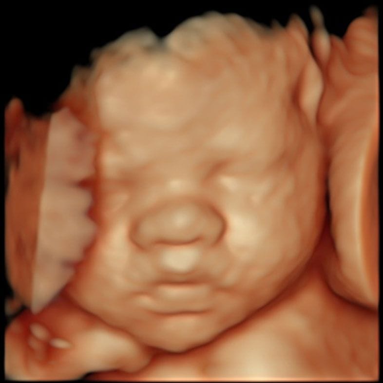
Third-trimester ultrasound image of a baby's abdomen during a growth assessment

Fetal growth restriction in numbers

1 in 10 By definition 1 in 10 babies measures below the 10th centile; most are simply small and healthy (RCOG)
3rd centile Below the 3rd centile, size alone defines growth restriction (Delphi consensus, adopted by ISUOG 2020)
30% / 70% About 30% of FGR is early-onset and 70% late-onset, split at 32 weeks (ISUOG, 2020)
71.6% Scan-estimated weight fell within 10% of actual birth weight in 71.6% of 11,049 term births (Dittkrist et al., 2022)
1.2% vs 1.7% Perinatal deaths in high-risk pregnancies with versus without umbilical artery Doppler-guided care, across 16 trials (Cochrane, 2017)
1 in 100 At term, babies below the 3rd centile carry the highest stillbirth risk — which is why birth is not left late (ISUOG, 2020)

How a small baby is picked up

Most pregnancies start with a tape measure. The midwife measures the bump from the top of the womb to the pubic bone — the symphysis-fundal height; the NHS schedule lists this from the 25-week visit, and RCOG guidance from 24 weeks. It is a screening step, not an answer: the measurement is affected by fibroids, the baby's position, fluid volume and maternal build.

If the measurement is small, or has flattened between visits, the next step is a growth scan — head, abdomen and thigh bone combined into an estimated fetal weight, plus the fluid and the blood flow in the cord and the baby's vessels. Many UK units plot the result on a customised chart adjusted for maternal height, weight, ethnicity and parity.

Scans estimate weight; they do not weigh. In 11,049 term deliveries the estimate fell within 10% of the actual birth weight 71.6% of the time, mean error 2.39% (Dittkrist et al., 2022) — close to three in ten are out by more than a tenth. That is why one measurement is rarely acted on alone. If a result does not fit the picture, a second opinion scan re-measures rather than repeats; where restriction is unexplained the team may look for a cause with NIPT or amniocentesis.

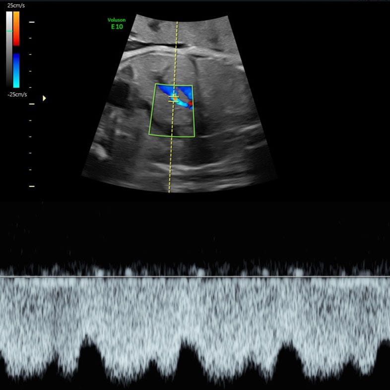
Doppler waveform showing blood flow in a fetal vessel during a growth assessment
Doppler studies show how well the placenta is keeping up

What a growth scan actually measures

Five things are looked at together, and it is the combination — not any single value — that separates a small healthy baby from one whose placenta is falling behind.

Estimated fetal weight (EFW)

Head circumference, abdominal circumference and femur length are combined into an estimated weight and plotted as a centile. This is the number everyone fixates on, and it carries a real margin of error — within 10% of actual birth weight about 71.6% of the time (Dittkrist et al., 2022). Read it as a position on a trend line.

Abdominal circumference (AC)

The tummy measurement is the most sensitive single marker of placental function: the liver and fat stores are the first things a baby stops investing in when supply tightens. An AC below the 3rd centile defines restriction on its own; below the 10th centile it counts when blood flow is also abnormal.

Umbilical artery Doppler

This measures resistance in the cord vessels — how hard your baby's heart must push to get blood through the placenta. A pulsatility index above the 95th centile suggests strain; absent or reversed end-diastolic flow needs immediate specialist care. In the 2017 Cochrane review, high-risk pregnancies with Doppler-guided care saw fewer perinatal deaths (1.2% versus 1.7%) across 16 trials and 10,225 babies.

Middle cerebral artery and CPR

The middle cerebral artery Doppler shows whether your baby is diverting blood to the brain — brain sparing. The cerebroplacental ratio (CPR) compares the two vessels; a CPR below the 5th centile is one criterion for late growth restriction. ISUOG calls these the most important Doppler measurements after 32 weeks — often where a baby of normal size turns out to need watching.

Fluid and growth velocity

A baby with a tired placenta passes less urine, so the fluid around them reduces; reduced liquor alongside a small measurement shifts the picture towards genuine restriction. Assessment may extend to a biophysical profile or computerised CTG.

Early-onset and late-onset growth restriction

Growth restriction behaves so differently before and after 32 weeks that specialists treat it as two conditions. ISUOG uses 32 weeks as the dividing line: about 30% of cases are early-onset, about 70% late-onset. In early FGR the difficulty is management — how long to keep a very premature baby inside a failing placenta. In late FGR the difficulty is detection.

Early-onset FGR is diagnosed when the AC or estimated weight is below the 3rd centile, or when there is absent end-diastolic flow in the umbilical artery; also when a measurement below the 10th centile combines with raised resistance in the umbilical or uterine arteries. It often accompanies pre-eclampsia, and monitoring happens in a fetal medicine unit.

Late-onset FGR, from 32 weeks, is subtler and more common. The criteria are an AC or estimated weight below the 3rd centile, or at least two of: a measurement below the 10th centile, growth crossing centiles by more than two quartiles, and a CPR below the 5th centile or umbilical artery resistance above the 95th centile. These babies can look normal on size alone — the abnormality often shows in the brain vessels instead.

Monitoring, and how the timing of birth is decided

How often scans are repeated

Growth scans are spaced at least two weeks apart, because the error in an estimated weight is larger than the growth expected over a shorter interval. Dopplers can be repeated far more often. NHS England's Saving Babies' Lives care bundle makes umbilical artery Doppler the primary surveillance tool for restriction found before 34+0 weeks, at minimum every two weeks.

What changes after 34 weeks

From 34 weeks the emphasis shifts to the brain vessels. ISUOG advises monitoring once or twice weekly where umbilical artery resistance is raised, noting that in one large study the median interval between a low middle cerebral artery pulsatility index and stillbirth after 34 weeks was five days or less. An abnormal result is confirmed within 24 hours before it is acted on.

Birth at 37 weeks

Where the estimated weight is below the 3rd centile, NHS England sets birth at 37+0 weeks and no later than 37+6. ISUOG agrees: babies below the 3rd centile at term carry the highest stillbirth risk, around 1 in 100. If birth is expected preterm, steroids mature the lungs: the RCOG offers them between 24+0 and 34+6 weeks, with the balance of benefit and risk weighed individually beyond 35+0.

Birth at 39 weeks

For a baby between the 3rd and 10th centiles with otherwise normal findings, the plan is later: birth at 39+0 weeks, achieved by 39+6. RCOG patient information frames it the same way — the latest you will be advised to give birth, if your baby is small but well, is between 39 weeks and your due date.

Where a private scan fits

A private growth scan can measure your baby, assess the Dopplers and give you a written report between NHS appointments. It does not replace your NHS pathway and it cannot change how your placenta works. What it gives you is measurements and blood-flow indices on the day, a specialist to explain them, and a report your maternity team can act on. Dr Fred Ushakov sees these cases directly.

Your questions answered

My baby is measuring small — should I be worried?

Usually not, and the numbers are on your side: one baby in ten measures below the 10th centile, and RCOG patient information is explicit that many babies are small and have no health issues. What decides whether it matters is the pattern rather than the single figure — whether your baby has always been on that line or has recently dropped away from it.

What is the difference between SGA and fetal growth restriction?

SGA describes size: the estimated weight or abdominal circumference is below the 10th centile. It says nothing about whether your baby is well. Fetal growth restriction describes a process — a pathological restriction of your baby's genetic growth potential, almost always because the placenta is underperforming. Many SGA babies are not restricted at all, and some restricted babies never drop below the 10th centile.

How accurate is a growth scan?

Less precise than most people assume. In 11,049 term deliveries the estimate came within 10% of the actual birth weight 71.6% of the time, mean error 2.39% (Dittkrist et al., 2022) — roughly three in ten are out by more than a tenth. That is why scans are spaced a fortnight apart and the trend is read rather than the single figure.

What is a Doppler scan and what does it show?

The same ultrasound machine used differently: instead of measuring size it measures how blood is moving. In the umbilical artery it shows how much resistance the placenta is putting up; in the middle cerebral artery, whether your baby is diverting blood to the brain. Together these answer the question size cannot — not how big is this baby, but how well is this baby being supplied.

What causes fetal growth restriction?

Usually the placenta. It may have implanted shallowly in early pregnancy, so that by the second half it cannot keep pace with a growing baby's demands. This is the same process that produces pre-eclampsia, which is why the two often travel together. Less common causes include cytomegalovirus, chromosomal differences and some structural anomalies. Smoking is the most significant modifiable factor.

Will my baby need to be born early?

Often earlier than 40 weeks, but for most families that means late term rather than premature. Below the 3rd centile, NHS England sets birth at 37+0 weeks and no later than 37+6. Between the 3rd and 10th centiles with normal blood flow, the plan is 39+0 weeks, by 39+6. Genuinely early birth is reserved for early-onset restriction where the Dopplers show the placenta failing.

Can anything be done to make my baby grow?

No treatment reliably makes a growth-restricted baby grow. Trials of drugs intended to improve placental blood flow, including sildenafil, have not shown benefit, and eating more does not change what a placenta can transfer. That is worth saying plainly, because many parents end up blaming themselves. Stopping smoking genuinely helps, at any stage. Beyond that, the value lies in monitoring rather than treatment.

What does it mean if my baby is crossing centiles?

It means the growth line has changed direction — your baby was tracking one centile and has dropped towards a lower one. This matters because it can happen while the baby is still, on paper, a normal size: one falling from the 60th centile to the 12th is still above the 10th. Crossing centiles by more than two quartiles is a consensus criterion for late growth restriction.

My bump measured small with a tape measure — does that mean my baby is small?

Not necessarily. Symphysis-fundal height is a screening step, not a measurement of your baby. It is affected by fluid volume, the baby's position, fibroids and your own build, and different people measure at different appointments. That is why the response to a small fundal height is a growth scan rather than a conclusion.

Is fetal growth restriction the same as IUGR?

Yes. IUGR — intrauterine growth restriction — is the older term for the same condition, and you will still see it in hospital notes and older leaflets. The preferred term now is fetal growth restriction, partly because the concern is the fetus not achieving its potential rather than the uterus.

Will my baby be all right?

For most babies who measure small, yes — they are small because that is who they are, and they are born well. That deserves to be said first. Where growth is genuinely restricted, it depends on how early it appears, how severe it is and how well your baby is coping; no scan and no clinician can promise you an outcome. Monitoring exists so the decision about when your baby is born is made deliberately. If you are frightened between appointments, contact your maternity unit — reduced movements always warrant a call the same day.

Contact

Speak to us about a growth scan

Send us an enquiry

A clinician will reply within one working day.

For urgent matters please call 020 3687 2939.

Sources & clinical references

The figures and clinical statements on this page are drawn from the sources below. Guidance evolves — always discuss your individual circumstances with a clinician.

  1. Royal College of Obstetricians and Gynaecologists (RCOG)Investigation and Care of a Small-for-Gestational-Age Fetus and a Growth Restricted Fetus (Green-top Guideline No. 31)2024
  2. Royal College of Obstetricians and Gynaecologists (RCOG)Having a small baby — patient information2024
  3. International Society of Ultrasound in Obstetrics and Gynecology (ISUOG)ISUOG Practice Guidelines: diagnosis and management of small-for-gestational-age fetus and fetal growth restriction2020
  4. NHS EnglandSaving babies' lives version 3: a care bundle for reducing perinatal mortality (Element 2 — fetal growth)2023
  5. Ultrasound in Obstetrics & Gynecology (Gordijn et al.), via PubMedConsensus definition of fetal growth restriction: a Delphi procedure2016
  6. Cochrane Database of Systematic Reviews (Alfirevic et al.), via PubMedFetal and umbilical Doppler ultrasound in high-risk pregnancies2017
  7. BMC Pregnancy and Childbirth (Dittkrist et al.)Percent error of ultrasound examination to estimate fetal weight at term in different categories of birth weight2022
  8. Tommy'sFetal growth restriction (intrauterine growth restriction)2024
  9. NHSYour antenatal appointments2024