
Echogenic Intracardiac Focus

A bright spot in your baby’s heart — often called a “golf ball” — is an echogenic intracardiac focus, or EIF. It is a speck of calcium in one of the tiny muscles inside the heart. It is not a hole, not a heart defect, and it does not change how the heart pumps. It is seen in about 4.6% of all pregnancies, roughly 1 in 22.

A bright spot is not a heart defect

If you have just been told there is a bright spot on your baby’s heart, the short answer comes first. An isolated echogenic intracardiac focus in a low-chance pregnancy is a normal variant. The Society for Maternal-Fetal Medicine calls it a normal variant of no clinical importance, with no indication for fetal echocardiography, follow-up imaging or any check after birth. The NHS Fetal Anomaly Screening Programme lists echogenic foci in the heart — alongside choroid plexus cysts and a two-vessel cord — as normal variants needing no referral.

What the sonographer sees is a small area that reflects sound as brightly as bone. It sits in or beside a papillary muscle — one of the tiny muscles anchoring the heart valve flaps — and is thought to be microcalcification or fibrosis: a speck of mineral in muscle tissue. It is not a hole between chambers and not a blocked valve, and studies using echocardiography and tissue Doppler have not found cardiac function affected by an isolated EIF.

It is usually temporary too, often fading in the third trimester or after birth. In a 2024 systematic review, 97% of babies in whom an EIF was seen had no cardiac abnormality at all. If you take one thing from this page, take that. For what a genuine structural problem looks like, see congenital heart defects.

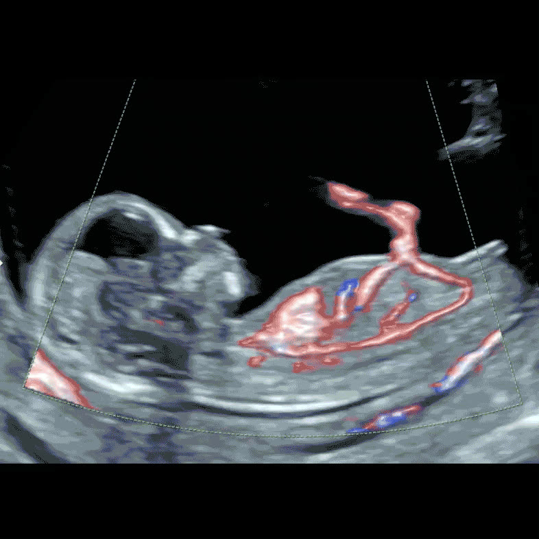
Colour Doppler ultrasound showing blood flow through a developing fetal heart

Echogenic intracardiac focus: the numbers

4.6% of all pregnancies show an echogenic intracardiac focus — about 1 in 22 (Jones et al., PLOS ONE systematic review, 2024)
5–10% is the prevalence usually quoted for a normal second-trimester population; individual studies range from 0.17% to 20% of pregnancies
4.2% of 8,207 consecutive scans at 14–23 weeks showed an EIF in babies with normal chromosomes (Borgida et al., 2005)
97% of babies in whom an EIF is seen have no cardiac abnormality of any kind (Jones et al., 2024)
86% of echogenic foci sit in the left ventricle; 3% are on the right and 10% are on both sides (Jones et al., 2024)
14.8% of 148 Japanese women in one series had a fetal EIF, against 7.3% of 5,948 pooled published controls (Rebarber et al., 2004)

Why anyone mentions it: the soft marker story

An EIF is what fetal medicine calls a soft marker — not an abnormality, but a normal-variant appearance that turned up somewhat more often in studies of babies with Down’s syndrome, in an era when ultrasound was one of the main ways of refining a woman’s chance. That is the entire reason a speck of calcium ever found its way into a scan report.

The numbers behind the reputation are weaker than the reputation. A meta-analysis of 48 studies put the pooled positive likelihood ratio for an echogenic intracardiac focus at 5.83 (95% CI 5.02–6.77), with a negative likelihood ratio of 0.

80 — but that draws heavily on referral populations already selected for higher chance, and those authors concluded most isolated markers produce only a small effect. In 44,103 low-risk pregnancies scanned at 18+0 to 21+6 weeks, an isolated echogenic focus was found in 5.05% and carried a positive likelihood ratio of roughly 2.3 to 3.1 for trisomy 21.

Sources disagree, so here is the range rather than the flattering end of it: the multiplier is in the low single digits at most, applied to a chance that was already very small.

What has changed is everything around the marker. NIPT — cell-free DNA screening from a sample of your blood — is far more informative about Down’s syndrome than any second-trimester appearance, so once a low-chance result is in hand a soft marker adds little. UK practice says so plainly: T21, T18 and T13 chance results must not be recalculated following the 20-week screening scan.

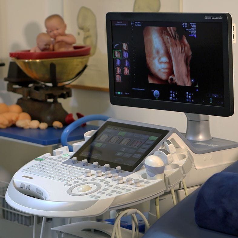
Ultrasound scanning equipment used for detailed fetal cardiac assessment
Equipment and operator variation affect how often a focus is reported

Isolated, or alongside something else?

Almost everything about how an echogenic intracardiac focus should be handled turns on one question: is it the only finding?

Most common

Isolated EIF, low-chance screening

The bright spot is the only thing seen, the anatomy looks normal, and your combined test or NIPT was low chance. Guidance here is unambiguous: no fetal echocardiogram, no repeat scan for the marker, no paediatric check after birth. The NHS anomaly-scan protocol requires no referral at all, and nothing about your care needs to change.

Isolated EIF, no screening yet

The bright spot is the only finding, but you have had no combined test or NIPT — so the marker is the only information anyone has. Guidance suggests counselling to estimate the chance of trisomy 21, with a discussion of options such as NIPT. That is a conversation about whether you want more information, not a step towards a procedure, and genetic counselling can help you decide. Declining is a legitimate choice.

EIF alongside other findings

The picture changes when the bright spot is not alone — an increased nuchal fold, ventriculomegaly, echogenic bowel, short long bones, or a structural difference in the heart itself. Combinations carry considerably more weight than any single marker, and the NHS programme lists several of those as needing referral in their own right. The next step is a specialist look at the whole anatomy, starting with an anomaly scan.

Right-sided or bilateral foci

Most foci — 86% — sit in the left ventricle. Right-sided foci are uncommon, at roughly 6 per 1,000 pregnancies, and carry a modestly higher association with cardiac abnormality: 7.9% in one systematic review, against 3.6% for left-sided foci. That justifies an unhurried look at the heart’s structure, not an assumption of a problem. Most right-sided foci are still found in normal hearts.

What the scan can and cannot tell you

An EIF is a mid-trimester observation. The studies defining it looked at scans between roughly 14 and 24 weeks — 14 to 23 weeks in one review of 8,207 pregnancies, 18+0 to 21+6 weeks in another of 44,103. That is the window in which the papillary muscles are clearly resolvable. An early dating or viability scan is not looking for it, and its absence earlier means nothing.

A scan can confirm whether the appearance really is a simple echogenic focus, whether it is single or bilateral, which side it is on and — far more usefully — whether the four chambers, outflow tracts and great vessels are structurally normal. An anomaly scan covers that as part of a full anatomical survey; a fetal cardiac scan or early fetal echocardiogram examines the heart in more detail. What no scan can do is make the finding go away or change the outcome for your baby.

How often an EIF gets reported also depends on who is scanning and on what. Discrepancies between studies may come down to differences in equipment and facilities, and identification has poor inter-rater reliability — when one study sent its suspected foci to a quality-assessment panel, 30% were not confirmed as an EIF at all. That is part of why published rates range from 0.17% to 20%, and part of why national guidance classes the finding as a normal variant.

If you left your appointment unsure what was examined, a second opinion scan with a fetal medicine specialist can work through the anatomy and explain what was and was not assessed. That is a way of understanding your scan, not a treatment — and if your finding is an isolated EIF with low-chance screening, guidance says you need no further imaging. We would rather tell you that than sell you a scan.

What usually happens next

Usually, nothing at all

For an isolated EIF with low-chance screening, the recommended management is no further evaluation: no fetal echo, no follow-up scan, no newborn cardiac check. If your midwife said it was nothing to worry about, she was following national guidance.

Screening, if you have not had it

Where no combined test or cell-free DNA screening has been done, the reasonable offer is screening rather than a procedure. NIPT uses a blood sample and says far more about trisomy 21 than any ultrasound marker.

A specialist second opinion

If you left your scan without a clear explanation, a fetal medicine review can go through the whole anatomy and say plainly what was seen. For an isolated EIF it is optional.

Diagnostic testing is not indicated

An isolated soft marker after low-chance screening is not a reason for amniocentesis or CVS. Invasive testing carries its own small risks, and guidance recommends against it for a soft marker alone.

Your questions answered

Is a bright spot in my baby’s heart serious?

On its own, no. An isolated echogenic intracardiac focus is a normal variant — not a structural problem, and no effect on how the heart pumps. In a 2024 systematic review, 97% of babies in whom an EIF was seen had no cardiac abnormality.

The NHS screening programme lists it among the normal variants that need no referral. If it was the only thing seen and your screening was low chance, there is nothing to act on.

What is the golf ball in my baby’s heart?

“Golf ball” is the informal phrase sonographers use, because the focus looks like a small bright spot on the screen. The medical name is echogenic intracardiac focus.

It is a small area of microcalcification or fibrosis in or beside a papillary muscle — one of the tiny muscles that tether the heart valve flaps. It reflects ultrasound as brightly as bone, which is why it stands out so sharply.

Does an echogenic intracardiac focus mean my baby has Down’s syndrome?

No. It is a soft marker — it was found somewhat more often in babies with Down’s syndrome in older studies, not that it indicates the condition. It is seen in about 4.6% of all pregnancies, and the overwhelming majority of those babies have normal chromosomes.

A meta-analysis of 48 studies gave a pooled positive likelihood ratio of 5.83 (95% CI 5.02–6.77) but concluded isolated markers produce only a small effect. In 44,103 low-risk pregnancies an isolated focus carried a likelihood ratio of roughly 2.3 to 3.1 — applied to a small starting chance, that still leaves a small chance.

Will the bright spot go away?

Usually. Echogenic foci are typically transient and often resolve in the third trimester or after delivery, so many are simply not visible on a later scan.

Whether it disappears or not does not change the significance. A focus still visible later in an otherwise normal heart is the same normal variant, still there.

Does an EIF mean my baby has a heart defect?

No. An EIF is not a heart defect, and studies using echocardiography and tissue Doppler have not found cardiac function affected by an isolated focus. In the published literature, 97% of babies in whom an EIF was detected had no cardiac abnormality.

Where a structural problem does exist, it is found by examining the chambers, valves and great vessels — see congenital heart defects — not by looking at the bright spot.

Will my baby need surgery or treatment?

No treatment exists for an echogenic intracardiac focus and none is needed. There is nothing to repair: it is a speck of calcium in muscle tissue, not a defect in the heart’s structure.

Guidance for an isolated EIF with a negative screening result states specifically that no evaluation after birth is indicated.

Do I need any more tests after an EIF is found?

If the focus is isolated and you have already had a low-chance combined test or NIPT, the recommendation is no further evaluation — no fetal echocardiogram, no follow-up imaging, no newborn check.

If you have had no screening at all, the reasonable offer is screening rather than a procedure: counselling about your chance of trisomy 21 and options such as NIPT. Amniocentesis is not recommended for an isolated soft marker after low-chance screening.

Why is an EIF more common in some ethnic groups?

It genuinely is. In one series, 14.8% of 148 Japanese women had a fetal EIF against 7.3% of 5,948 pooled published controls; a study cited in that paper reported 30.4% among a small group of 46 Asian patients. A review of 8,207 pregnancies with normal chromosomes found an overall rate of 4.2%, and babies of Asian mothers were significantly more likely to have one.

Nobody knows why. The consequence is that the finding means even less where it is common — the more often a marker appears in unaffected pregnancies, the less it says about any individual one. These samples are small, so treat the higher percentages as a range.

My NIPT was low chance — does the bright spot change that?

In practical terms, no. Guidance is explicit that an isolated soft marker after a negative cell-free DNA or serum screening result is not a reason for diagnostic testing, and UK practice reinforces it: T21, T18 and T13 chance results must not be recalculated following the 20-week scan.

NIPT remains a screening test rather than a diagnostic one, so a low-chance result is strong reassurance rather than certainty — but a bright spot does not weaken it.

Does it matter which side of the heart the spot is on?

Slightly. About 86% of echogenic foci are in the left ventricle, 3% on the right and 10% on both sides; right-sided foci are uncommon, at roughly 6 per 1,000 pregnancies.

In one systematic review, 7.9% of right-sided foci were associated with a cardiac abnormality, against 3.6% of left-sided foci. That warrants a careful structural look at the heart — a cardiac scan or fetal echocardiogram — rather than an expectation of a problem.

Should I book a second opinion scan?

Only if you want one, and not because of the marker itself. For an isolated EIF with low-chance screening, national guidance is that no further imaging is indicated, and we would rather say so plainly than imply otherwise.

A second opinion scan helps if you left your appointment without a clear explanation, if you are unsure whether the heart and the rest of the anatomy were fully assessed, or if the focus was right-sided or bilateral. A review with Dr Fred Ushakov can go through what was seen and what it means — it cannot change the finding or promise reassurance.

Contact

Talk to us about a finding on your scan

Send us an enquiry

A clinician will reply within one working day.

For urgent matters please call 020 3687 2939.

Sources & clinical references

The figures and clinical statements on this page are drawn from the sources below. Guidance evolves — always discuss your individual circumstances with a clinician.

  1. NHS England / GOV.UK — Fetal Anomaly Screening Programme (FASP) handbook20-week screening scan2026
  2. Society for Maternal-Fetal Medicine (SMFM)Consult Series #57: Evaluation and management of isolated soft ultrasound markers for aneuploidy in the second trimester2021
  3. PLOS ONE (Jones HE, Battaglia S, Hurt L, Uzun O, Brophy S)Echogenic intracardiac foci detection and location in the second-trimester ultrasound and association with fetal outcomes: a systematic literature review2024
  4. Ultrasound International Open (Wrede E, Knippel AJ, Verde PE, Hammer R, Kozlowski P)Isolated echogenic cardiac focus: assessing association with trisomy 21 by combining results from a prenatal center with a Bayesian meta-analysis2020
  5. Ultrasound in Obstetrics & Gynecology (Agathokleous M, Chaveeva P, Poon LCY, Kosinski P, Nicolaides KH)Meta-analysis of second-trimester markers for trisomy 212013
  6. Journal of Maternal-Fetal & Neonatal Medicine (Borgida AF, Maffeo C, Gianferarri EA, Bolnick AD, Zelop CM, Egan JFX)Frequency of echogenic intracardiac focus by race/ethnicity in euploid fetuses2005
  7. BMC Pregnancy and Childbirth (Rebarber A et al.)An ethnic predilection for fetal echogenic intracardiac focus identified during targeted midtrimester ultrasound examination: a retrospective review2004
  8. NHSScreening for Down's syndrome, Edwards' syndrome and Patau's syndrome2024