
Chromosomal conditions
Exomphalos is associated with trisomy 18 (Edwards' syndrome), trisomy 13 (Patau's syndrome) and trisomy 21 (Down's syndrome). This is why genetic testing is usually offered when exomphalos is found.
Exomphalos — also called an omphalocele — is an abdominal wall defect in which some of the baby's abdominal organs develop outside the tummy at the base of the umbilical cord, protected by a thin membrane. It affects around 1 in 3,000 pregnancies and can often be seen on ultrasound from the first trimester.
Exomphalos is an abdominal wall defect — see also body stalk anomaly and cloacal exstrophy, related conditions detectable early in pregnancy.
Exomphalos happens when the muscles of the baby's abdominal wall do not close fully during early development, leaving a gap at the base of the umbilical cord. Abdominal organs then pass through this gap and sit outside the body, enclosed in a protective sac formed by the membranes that normally line the abdomen and cord.
When only the bowel is involved, the defect is usually described as minor (exomphalos minor). When the liver is also outside the abdomen — the 'exomphalos with liver' picture — it is described as major (exomphalos major). The covering membrane is what distinguishes exomphalos from gastroschisis, a separate abdominal wall defect in which the bowel is exposed without any covering.
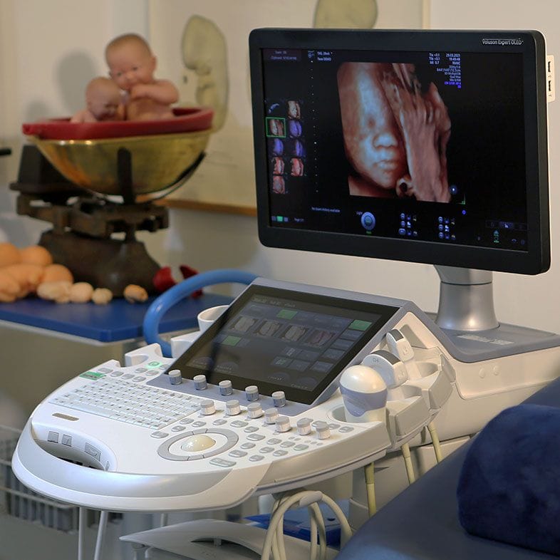
Exomphalos can often be identified on ultrasound. In early pregnancy the bowel normally herniates into the base of the cord and returns to the abdomen by about 12 weeks, so a confident diagnosis is usually made after this point — frequently at an early fetal or 11–14 week scan, and confirmed at the 20-week anomaly scan.
Because exomphalos is strongly associated with chromosomal and heart conditions, finding it prompts a detailed structural scan, a fetal heart scan (echocardiography), and a discussion about genetic testing such as NIPT, chorionic villus sampling (CVS) or amniocentesis. Identifying the defect early gives parents time for these investigations and to plan care and delivery with a specialist team.
At London Pregnancy Clinic our early scans are performed by fetal medicine specialists who routinely assess the abdominal wall as part of a structured examination. If exomphalos is suspected, we can arrange a detailed follow-up scan, assess the size of the defect and whether the liver is involved, and look carefully for other features.
Because of the strong link with chromosomal conditions, we can discuss and arrange NIPT and, where appropriate, diagnostic testing, alongside referral for a fetal echocardiogram. Our Fetal Medicine and Obstetrics consultant, Ms Diane Nzelu, is available for consultations to guide you through the findings, the options and what they mean for your pregnancy.

Exomphalos is often isolated, but it can occur alongside chromosomal, genetic or structural conditions. When exomphalos is found, careful assessment for the following is important:
Exomphalos is associated with trisomy 18 (Edwards' syndrome), trisomy 13 (Patau's syndrome) and trisomy 21 (Down's syndrome). This is why genetic testing is usually offered when exomphalos is found.
An overgrowth condition that is one of the more common syndromes linked with exomphalos, sometimes associated with a large tongue, low blood sugar in the newborn period and increased birth weight.
Heart abnormalities are among the most frequent associated findings, which is why a fetal heart scan (echocardiography) is recommended when exomphalos is diagnosed.
A rare combination of midline defects that can include an abdominal wall defect, a diaphragm defect and heart abnormalities.
Related midline problems such as cloacal exstrophy and bladder or bowel abnormalities may occasionally be seen.
In many babies exomphalos occurs on its own, with no chromosomal or structural association — and here the outlook after surgery is generally very good.
Exomphalos, also called an omphalocele, is a defect in the front wall of the baby's tummy. During normal development the abdominal wall closes and the organs sit inside the abdomen. In exomphalos the muscles do not close fully, leaving a gap at the base of the umbilical cord through which abdominal organs — the bowel and, in larger cases, the liver — protrude. These organs are contained within a thin, protective membrane rather than being exposed.
Exomphalos is one of the two main abdominal wall defects, alongside gastroschisis, and affects around 1 in 3,000 pregnancies. It can range from minor (a small defect containing only bowel) to major (a larger defect that also contains the liver).
When the defect is large enough that the liver — as well as the bowel — lies outside the abdomen, it is described as major exomphalos, or 'exomphalos with liver'. Major exomphalos is a bigger defect and usually needs a staged surgical repair after birth, because the abdominal cavity may be too small to return all of the organs in a single operation.
The involvement of the liver is one of several factors, alongside the baby's overall health and any associated conditions, that specialists consider when planning delivery and treatment.
Both are abdominal wall defects in which organs develop outside the tummy, but they are distinct conditions. In exomphalos the organs herniate through the base of the umbilical cord and are covered by a protective membrane. In gastroschisis the defect is usually to the side of the umbilical cord and the bowel is exposed directly to the amniotic fluid, with no covering membrane.
The covering membrane, the position of the defect and the different pattern of associated conditions help specialists tell the two apart on ultrasound.
In early pregnancy the bowel normally moves into the base of the umbilical cord and returns to the abdomen by about 12 weeks. Because of this normal process, a confident diagnosis of exomphalos is usually made after 12 weeks — often at an early fetal or 11–14 week scan, and confirmed at the 20-week anomaly scan.
At London Pregnancy Clinic our early scans include a structured look at the abdominal wall, so suspicion can be raised early and a detailed follow-up arranged.
Exomphalos can occur on its own, but it is also associated with chromosomal conditions such as trisomy 18, 13 and 21, with Beckwith-Wiedemann syndrome, and with heart defects. When exomphalos is isolated the chromosomal risk is low (around 4–5%, roughly 1 in 20), but it is much higher (about 30–70%) when other abnormalities are present.
For this reason, when exomphalos is found, parents are usually offered detailed scanning, a fetal heart scan and genetic testing such as NIPT or a diagnostic test (CVS or amniocentesis) to give the fullest possible picture.
After birth the protective membrane is kept intact and the baby is kept warm and hydrated before transfer to a specialist surgical centre. Small defects can often be repaired in a single operation, in which the organs are returned to the abdomen and the abdominal wall is closed.
Larger defects, including those involving the liver, are usually treated in stages: a protective covering (sometimes called a silo) is placed over the organs and gradually tightened over days to weeks, allowing the abdomen to grow before final closure. Occasionally, if closure is not immediately safe, a patch is left in place and treated later.
The outlook depends largely on the size of the defect and whether there are any associated conditions. For isolated exomphalos — where the baby has no other problems — more than 95% of babies make a full recovery.
When exomphalos occurs alongside a chromosomal condition, a heart defect, a very large defect or prematurity, the outlook is more variable. Early diagnosis and assessment allow the specialist team to plan the safest possible care and to prepare parents for what to expect.
These organisations provide trusted information and support for parents of a baby diagnosed with exomphalos.
Official NHS screening information explaining what exomphalos is, how it is found, and what happens next, including the difference between minor and major exomphalos.
A leading UK children's hospital's guide to exomphalos, covering diagnosis, the protective membrane, and how the condition is repaired with surgery.
A UK charity offering non-directive information and support to parents through antenatal screening and diagnosis, including when a condition such as exomphalos is found.
Contact
The figures and clinical statements on this page are drawn from the sources below. Guidance evolves — always discuss your individual circumstances with a clinician.
Cookies on this site
We use cookies needed to run this site. With your permission we would also use analytics and advertising cookies to see how the site is used and which of our adverts lead to bookings. Declining will not affect your care or your booking. See our Privacy notice and Terms & Conditions.