
Exomphalos (omphalocele)
The other main abdominal wall defect. Here the organs stay covered by a membrane at the base of the umbilical cord and there is a higher chance of chromosomal or other structural problems, so the two are managed differently.
Gastroschisis is an abdominal wall defect in which the baby's bowel develops outside the tummy, protruding through a small opening usually to the right of the umbilical cord and, unlike exomphalos, not covered by a protective membrane. It is one of the 11 physical conditions the NHS Fetal Anomaly Screening Programme (FASP) looks for at the 20-week scan. At London Pregnancy Clinic our detailed 10 Week Scan and Early Fetal Scan, carried out by fetal-medicine specialists, can often raise suspicion of an abdominal wall defect well before 20 weeks.
Gastroschisis is one of the 11 conditions the NHS screens for at the 20-week scan — see our FASP overview and how we aim to detect anomalies earlier.
Gastroschisis happens when a baby's abdominal (tummy) wall does not close fully in early pregnancy, leaving a small gap. Loops of bowel, and occasionally other organs, pass through this opening and float freely in the amniotic fluid. The defect sits alongside the belly button, almost always to the right of it, and the exposed bowel has no covering membrane. This absence of a membrane is the key feature that distinguishes gastroschisis from exomphalos (omphalocele), where the organs remain enclosed within a sac at the base of the umbilical cord.
The exact cause is not known. Importantly, gastroschisis is usually an isolated finding: unlike many other structural anomalies, it is rarely linked to a chromosomal problem, and only around 10% of babies have an additional malformation outside the digestive system. It is one of the more common abdominal wall defects seen in pregnancy, affecting roughly 1 in 3,000 babies, and it is becoming more common in the UK, particularly among mothers under the age of 20.
Because the bowel is bathed in amniotic fluid throughout pregnancy, it can become swollen, thickened and irritated, and babies are often smaller than expected and born a little early. The outlook, however, is generally very good. Gastroschisis needs an operation shortly after birth to return the bowel to the abdomen, usually followed by a period of neonatal care, and around 9 in 10 babies make a full recovery.
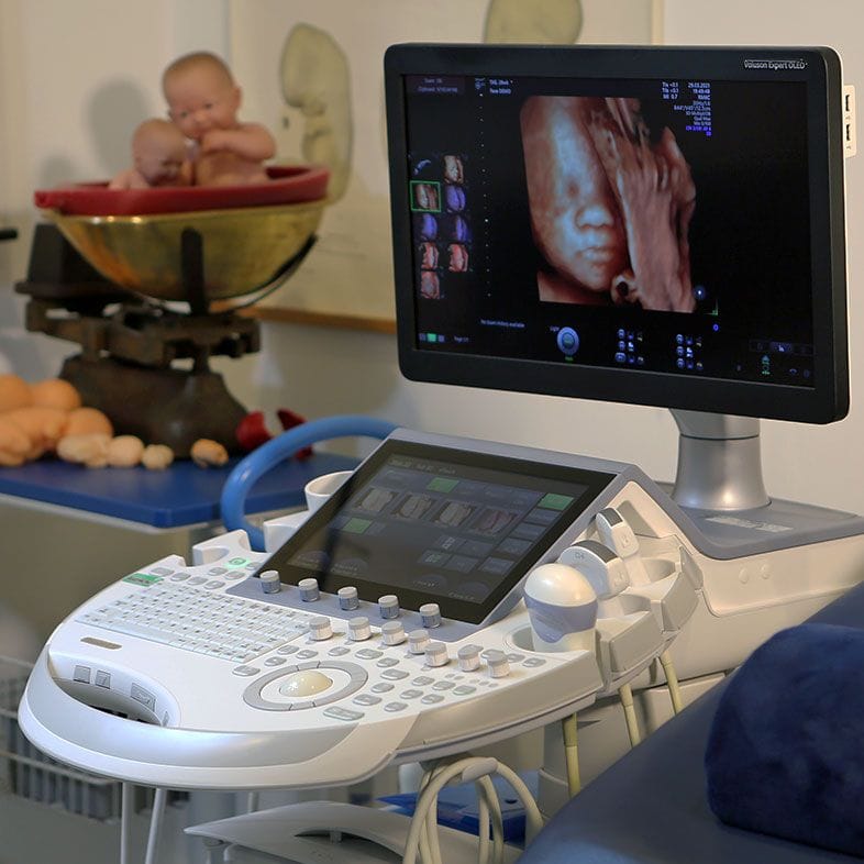
Gastroschisis is detected on ultrasound, where the sonographer sees loops of bowel floating freely in the amniotic fluid outside the abdomen, without a surrounding membrane. Maternal serum alpha-fetoprotein (AFP) is also typically raised. In the NHS pathway it is one of the 11 conditions specifically screened for at the 20-week scan (carried out between 18+0 and 20+6 weeks), and it is sometimes first noticed at the routine scan around 12 weeks. A confirmed diagnosis usually leads to referral to a fetal-medicine unit and planning for delivery at a hospital with neonatal surgery on site.
At London Pregnancy Clinic our emphasis is on earlier, more detailed assessment. Because gastroschisis involves bowel visibly outside a clear abdominal outline, it is one of the anomalies that can often be suspected before the 20-week scan.
Our detailed 10 Week Scan and Early Fetal Scan, performed by fetal-medicine specialists using high-resolution equipment, examine the developing abdominal wall closely and can frequently raise suspicion in the late first or early second trimester.
We are careful not to overpromise: the abdominal wall is still forming very early in pregnancy and some findings can only be confirmed reliably as the baby grows, so an early scan complements rather than replaces the 20-week anomaly scan. Where we do see something, an early answer means more time to plan, to arrange specialist referral and to prepare for your baby's care.
The other main abdominal wall defect. Here the organs stay covered by a membrane at the base of the umbilical cord and there is a higher chance of chromosomal or other structural problems, so the two are managed differently.
Because the exposed bowel is irritated by amniotic fluid, additional bowel problems occur in up to about a quarter of cases; a gap in the bowel (atresia) is less common but may need further surgery.
Babies with gastroschisis are often smaller than expected for their age, so growth is monitored closely with serial scans in the second and third trimesters.
Babies with gastroschisis are commonly born a little early, which is one reason delivery is planned at a hospital with neonatal and surgical care available.
In contrast to many anomalies, gastroschisis is rarely associated with a chromosomal condition, and only around 10% of babies have a malformation elsewhere in the body.
Gastroschisis is strongly associated with younger mothers and its incidence in the UK has risen over recent decades, most markedly among women under 20.
Both are abdominal wall defects, but the key difference is the covering. In gastroschisis the bowel passes through an opening beside the belly button (usually to the right) and is not covered by any membrane, floating freely in the amniotic fluid. In exomphalos the organs remain enclosed within a protective sac at the base of the umbilical cord.
This distinction matters clinically. Exomphalos carries a higher chance of chromosomal and other structural problems, whereas gastroschisis is usually an isolated condition. Your specialist will explain which one has been seen and what it means for your pregnancy.
Usually not. Gastroschisis is typically an isolated finding and is rarely associated with a chromosomal abnormality. Only around 10% of babies have an additional malformation outside the digestive tract.
This is reassuring, but your team may still discuss detailed scanning and, in some cases, further testing to look at the rest of the baby's development before making a full plan.
Often, yes. Gastroschisis is one of the abdominal wall defects that can be suspected earlier than 20 weeks because bowel is visibly outside a clear abdominal outline. It is sometimes first noticed at the routine scan around 12 weeks.
At London Pregnancy Clinic our detailed 10 Week Scan and Early Fetal Scan, performed by fetal-medicine specialists, look closely at the developing abdominal wall and can frequently raise suspicion earlier. Because the abdomen is still forming very early on, an early scan complements the 20-week NHS anomaly scan rather than replacing it.
On ultrasound the sonographer sees loops of bowel floating freely in the amniotic fluid outside the tummy, without a membrane around them. A raised level of a blood marker called alpha-fetoprotein (AFP) in the mother can also point towards the diagnosis.
If gastroschisis is confirmed you will usually be referred to a fetal-medicine unit, and delivery will be planned at a hospital with neonatal surgery available.
Immediately after birth the exposed bowel is gently wrapped to keep it warm and protected, and the baby is cared for by a specialist surgical team. An operation is then needed to return the bowel to the abdomen and close the wall.
This may be done in a single operation, or in stages using a protective sac (a silo or mesh) that gradually eases the bowel back inside over several days. Babies then usually spend a period in neonatal care while their bowel starts to work and feeding is established.
The outlook is generally very good. Around 9 in 10 babies with gastroschisis make a full recovery, and most grow up to live normal lives.
Recovery time varies. Babies with straightforward (simple) gastroschisis often spend around a month in hospital, while those whose bowel has been more affected may need longer care. Your surgical team will give you a clearer picture based on your baby's individual situation.
The incidence of gastroschisis has risen in the UK over recent decades, and it is most strongly associated with younger mothers, particularly those under the age of 20. The exact reasons are not fully understood.
What is clear is that this is a demographic association rather than something a parent has done. Your team will focus on monitoring and planning the safest possible care for you and your baby.
These organisations provide trusted information and support relating to gastroschisis.
The UK charity for babies born premature or sick. Bliss offers free emotional and practical support to families facing neonatal care, including an online community, Bliss Champion volunteers on neonatal units, and guidance on the neonatal journey.
A non-profit dedicated to research, awareness and support for families affected by gastroschisis, offering shared stories and information for parents (international, with resources relevant to UK families).
An NHS patient-information page explaining gastroschisis, its treatment and what to expect, with signposting to further support organisations.
Contact
The figures and clinical statements on this page are drawn from the sources below. Guidance evolves — always discuss your individual circumstances with a clinician.
Cookies on this site
We use cookies needed to run this site. With your permission we would also use analytics and advertising cookies to see how the site is used and which of our adverts lead to bookings. Declining will not affect your care or your booking. See our Privacy notice and Terms & Conditions.