
Pregnancy Stopped Growing: What Can Cause It?

Hearing that a pregnancy is measuring small, or may have stopped growing, is hard to take in, and one scan often cannot give a final answer. Our fetal medicine team explains how growth is checked over time, why development sometimes stops, and what can be tested once the picture is clear.

If you have heavy bleeding, severe pain or feel faint, call 999 or go to A&E. Otherwise your NHS early pregnancy unit can assess you and arrange a repeat scan free of charge.

Available atLPC City (E1)LPC West (SW10)

Understanding the scan

Measuring small is not the same as stopped growing

On an early scan, the clinician looks for a heartbeat first. If an embryo is visible, they measure its length from head to bottom, called the crown–rump length (CRL); if not, they measure the pregnancy sac. NICE, which sets guidance for the NHS, recommends this order, using an internal (transvaginal) scan where possible.

The measurements are compared with how far along you are thought to be, usually counted from your last period. Cycle lengths vary, so a pregnancy can be younger than the calendar suggests, and NICE advises that period dates alone should not decide whether a heartbeat ought to be visible yet.

So a small measurement can mean the pregnancy is earlier than expected, developing more slowly, or has stopped. If a heartbeat is seen, the pregnancy had not stopped developing at the time of the scan. 'Stopped growing' only fits when there is no heartbeat and miscarriage has been confirmed using NICE's criteria, which always involve a second opinion, a repeat scan or both.

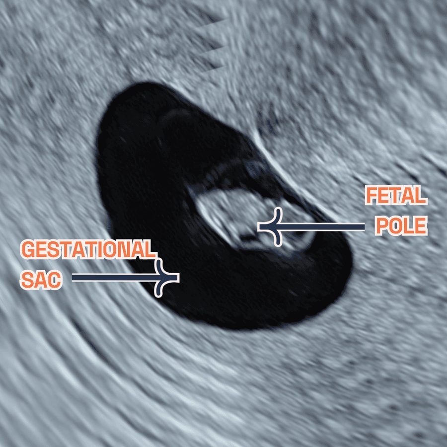
Early pregnancy ultrasound with the gestational sac and the fetal pole labelled
The fetal pole (early embryo) is measured head to bottom; the sac is measured if no embryo is seen

Three possibilities

What a small measurement can mean

Scan reports use different words for these situations. Follow-up scanning is how they are told apart.

Heartbeat seen

Earlier than your dates

The embryo is shorter than expected for your dates, but its heart is beating. The pregnancy may simply be younger than you thought, and a later scan can re-date it. Your clinician may still suggest a follow-up scan to check growth.

Too early to tell

Uncertain viability

A sac with no embryo yet, or a small embryo with no visible heartbeat, below the sizes NICE uses when diagnosing miscarriage. Doctors describe this as a 'pregnancy of uncertain viability'. The safe next step is another scan at least 7 days later.

Miscarriage confirmed

Development has stopped

No heartbeat once the embryo is large enough for one to be expected, confirmed by a second opinion and/or a repeat scan, or repeat scanning shows none has developed. This is a missed miscarriage. If the sac never held an embryo, see an empty pregnancy sac.

How it is assessed

How growth is checked over time

1

The first scan

An internal scan looks for a heartbeat, then measures the embryo or, if none is visible, the sac. NICE asks clinicians to explain that one scan cannot be 100% accurate, particularly very early on.

2

Checking against NICE size limits

If the embryo measures 7 mm or longer without a heartbeat, or an empty sac is 25 mm or wider, NICE recommends a second opinion and/or another scan a minimum of 7 days on, before miscarriage is diagnosed.

3

Below those sizes, waiting is essential

A smaller embryo with no heartbeat, or a smaller empty sac, always needs another scan no sooner than 7 days later, sometimes more than one. If only an abdominal scan was possible, NICE asks for at least 14 days between scans, whatever the measurements. NICE says waiting does not harm the pregnancy.

4

The repeat scan

This looks for a heartbeat not seen before and compares measurements. In a study of uncertain early pregnancies at four London hospitals, the embryo lengthened by roughly 0.67 mm a day on average when the pregnancy went on to continue, against about 0.15 mm a day when it miscarried.

5

Why growth is not judged on its own

Growth rates overlap between pregnancies that continue and those that do not, especially for the sac, so the researchers judged a diagnosis based on growth alone potentially unsafe. The heartbeat and NICE's size limits come first.

Uncertain early scans: the numbers

Figures from published studies describe groups of people, not any one pregnancy. Sources are listed at the foot of this page.

473 of 1,060 Uncertain early pregnancies at four London hospitals still continuing at the 11–14 week scan; the other 587 did not continue (Abdallah et al., 2011)
86 in 100 Pregnancies with a heartbeat at 5–10 weeks still continuing at 11–14 weeks in one early pregnancy unit; those that miscarried had measured smaller (Mukri et al., 2008)
About 1 in 2 Early miscarriages caused by a chromosome abnormality in the pregnancy (RCOG Green-top Guideline, 2023)

Causes

Why does a pregnancy stop developing?

Most of the time, nothing you did or did not do caused it. The most common reason is a chromosome abnormality in the pregnancy itself. Chromosomes carry the instructions for development, and an egg and a sperm should each bring one copy of every chromosome. If the egg or sperm carries an extra or missing chromosome, or the fertilised egg ends up with a whole extra set, development often stops in the first weeks.

The RCOG estimates that about half of early miscarriages happen this way. In a 2023 study from China of tissue from 1,160 early missed miscarriages, where development had stopped but the pregnancy had not come away, around 6 in 10 had a chromosome abnormality on genome-wide chromosome tests, not counting findings of uncertain meaning.

Most of these changes arise by chance as the egg or sperm forms, or in the first cell divisions, and the parents' own chromosomes are usually normal. Egg errors become more common with age as the number and quality of eggs decline, one reason miscarriage becomes more common as women get older.

A 2003 study used a fine camera to look at 233 embryos from missed miscarriages before surgery. About 3 in 4 of those tested had a chromosome abnormality, nearly 1 in 5 had a visible developmental problem despite normal chromosomes, and in about 7 in 100 no problem of either kind was found with the tests available at the time.

Chromosome changes

The chromosome changes found most often

When a change is found in miscarriage tissue, it is usually one of these. Our guide to chromosomal causes of miscarriage goes into more depth.

An extra chromosome (trisomy)

The most common finding: about half of abnormal results. Trisomy 16 is the most frequent single one, around a quarter of trisomies in one large study. Trisomies become more common as the egg gets older.

Extra whole sets (polyploidy)

Whole extra sets, such as three instead of two (triploidy): nearly 1 in 5 abnormal results. If the extra set comes from the father it can be a partial molar pregnancy, which needs hCG hormone follow-up until levels return to normal.

A missing chromosome (monosomy)

Usually monosomy X: one X chromosome instead of two sex chromosomes. Monosomies make up about 15 in 100 abnormal results, and monosomy X does not become more common with the mother's age.

Missing or extra pieces (structural)

Part of a chromosome is missing, duplicated or rearranged: about 6 or 7 in 100 abnormal results. Occasionally a parent carries a balanced rearrangement, which is when parental chromosome testing becomes relevant.

Beyond chromosomes

Other biological reasons development can stop

When the chromosomes are normal, a cause is harder to pin down, and sometimes none is found. Possibilities include:

  • Structural problems in the embryo: in the camera study above, nearly 1 in 5 embryos had a visible developmental problem even though their chromosome test, using the methods of the time, was normal.
  • Single-gene changes: too small for chromosome tests to see. A 2025 genome study estimated that around 1 in 136 pregnancies is lost because of one.
  • Tiny missing or extra pieces: a whole-genome test called a microarray finds these in a further 5 to 7 in 100 miscarriages, though not all explain the loss.
  • Health conditions in the mother: diabetes with high blood sugar in early pregnancy, thyroid disease, antiphospholipid syndrome (an immune condition that makes blood more likely to clot) and some differences in womb shape. Most are looked for after recurrent miscarriage.
  • Not everyday life: exercise, sex, ordinary work and everyday lifting do not cause miscarriage. See why did I miscarry?

Genetic testing

What can be tested, and when

Genetic tests are not used to decide whether a pregnancy that is measuring small will continue; scans over time answer that. Testing becomes relevant once a miscarriage is confirmed, if you would like to understand why.

Timing matters. Without treatment, a missed miscarriage often stays in the womb for days to weeks: in one UK study, about 3 in 10 had passed within a week and 6 in 10 within two weeks. While the pregnancy remains in the womb, a blood-based genetic test that reads placental DNA circulating in your blood may be possible; see testing before a miscarriage completes.

A blood-based result is a screening-type answer, not a diagnosis. Across eight studies it found nearly 4 in 5 of the chromosome changes that tissue testing found; the DNA-counting method used in all of those studies cannot detect triploidy, and some samples give no result. If you have a procedure or collect tissue, pregnancy tissue testing examines the chromosomes directly.

On the NHS, tissue testing is usually offered from the third miscarriage, or after any second-trimester miscarriage. After a first or second loss, our genetic counsellors can help you decide whether a private test is possible and worthwhile. For a step-by-step guide, see what can I test now?

Where you are now, and what to discuss

A simplified guide. Your own history and scan findings may change the advice you are given.

Where you are nowWhat usually happens nextGenetic testing to discuss
Measuring small, heartbeat seenA follow-up scan if your clinician advises oneNone at this stage; the pregnancy is ongoing
No heartbeat yet, below NICE sizesAnother scan after a gap of at least 7 days (14 days if abdominal only)None yet; wait for a clear diagnosis
Miscarriage confirmed, pregnancy still in the wombA decision about how the miscarriage is managed; see missed miscarriageA blood-based test before treatment, or tissue testing afterwards if tissue can be collected
Pregnancy tissue passed or removedRecovery and follow-up careTissue testing if suitable tissue is available
Repeated losses, nothing tested yetInvestigations for recurrent miscarriagePossibly parental chromosome tests

At London Pregnancy Clinic

Our scans and genetics support

Your NHS early pregnancy unit can assess you and arrange repeat scans free of charge, and for many people that is all that is needed. We can help if you would like an independent opinion, a convenient appointment or advice on genetic testing. A private scan cannot shorten the 7-day gap NICE recommends between scans.

6–9 weeks

Viability scan

A repeat scan by our fetal medicine doctors and specialist sonographers to look for a heartbeat and growth, best booked at least 7 days after your previous scan.

Uncertain findings

Second-opinion scan

From around 10 weeks, a detailed scan with Dr Fred Ushakov, who has a particular interest in the earliest stages of pregnancy, for an independent view of an uncertain or upsetting result.

Online, 30 minutes

Genetic counselling

A video appointment with a registered genetic counsellor, through our partner Jeen Health, to discuss whether testing is possible now and which test fits.

Questions about a pregnancy that has stopped growing

My scan says the pregnancy is measuring a week behind. Will it miscarry?

Not necessarily. If a heartbeat was seen, the pregnancy is developing and may simply have started later than your dates suggest, especially if your cycles vary. In one study of pregnancies with certain dates, embryos that continued still measured slightly smaller than the reference chart on average, but those that later miscarried had measured clearly smaller. So measuring small does carry a higher chance of miscarriage, which is why a follow-up scan may be suggested.

Why do I have to wait at least a week for the next scan?

Because real change takes time to show. A heartbeat too early to see today may be clear a week later, and embryos in continuing pregnancies grow by less than a millimetre a day on average. NICE recommends at least 7 days between scans and says the wait does not harm the pregnancy. If you develop heavy bleeding or pain meanwhile, get seen straight away.

Can a pregnancy that seemed to have stopped growing turn out to be fine?

Yes, if the first scan was too early to be sure, which is why NICE asks for a repeat scan first. In the London study above, 473 of 1,060 pregnancies of uncertain viability were still continuing at 11–14 weeks, while the other 587 were not, so the repeat scan matters either way. Once miscarriage has been confirmed using NICE's criteria, though, development does not restart.

Could exercise, work or stress have stopped the pregnancy growing?

This is very unlikely. The most common cause is a chance chromosome change present from the very start. Exercise, sex, ordinary work and everyday lifting do not cause miscarriage, and for everyday stress, research has found no clear evidence that it causes miscarriage. Smoking, alcohol (especially heavier drinking) and a lot of caffeine are linked with a higher risk, but a drink before you knew you were pregnant is very unlikely to explain it.

Should I have the repeat scan on the NHS or privately?

Your NHS early pregnancy unit will usually book it free of charge, which suits most people. Some choose a private scan for an independent opinion, a convenient time or more time to ask questions. Either way, book it at least 7 days after your last scan. Call us on 020 3687 2939 if you are unsure.

What if the scan shows a sac but no embryo?

Early on, a sac can be seen before the embryo, so one scan showing an empty sac is not enough on its own. Under 25 mm, NICE asks for another scan no sooner than 7 days later; at 25 mm or more, a second opinion and/or a repeat scan. A London study found that if the sac is still empty on two scans more than 7 days apart, miscarriage is highly likely, whatever the growth. Our guide to an empty pregnancy sac explains more.

Can a scan show why development stopped?

Not usually. A scan shows the heartbeat and the size of the embryo or sac, and occasionally features that suggest a molar pregnancy, but it cannot show the pregnancy's chromosomes. Only a genetic test of placental DNA in your blood, or of the pregnancy tissue, can look for a chromosomal cause, and even then a cause is not always found.

Support while you wait

The days between scans can be among the hardest. Our miscarriage support page lists charities and helplines, and you are welcome to call us on 020 3687 2939 to talk something through.

About this information

This page gives general medical information and cannot replace advice from a clinician who knows your history and has seen your scans. If you have heavy bleeding, severe pain, a fever or feel faint, contact your early pregnancy unit or call NHS 111 straight away, or call 999 in an emergency.

Contact

Questions about an uncertain scan?

Send us an enquiry

Not for emergencies: if you are bleeding heavily or in severe pain, call 999 or go to A&E.

For urgent matters please call 020 3687 2939.

Sources & clinical references

The figures and clinical statements on this page are drawn from the sources below. Guidance evolves — always discuss your individual circumstances with a clinician.

  1. NICEEctopic pregnancy and miscarriage (NG126): diagnosis of viable intrauterine pregnancy, recommendations 1.5–1.62026
  2. Ultrasound in Obstetrics & Gynecology (Abdallah et al.)Limitations of current definitions of miscarriage using mean gestational sac diameter and crown-rump length measurements: a multicenter observational study2011
  3. Ultrasound in Obstetrics & Gynecology (Abdallah et al.)Gestational sac and embryonic growth are not useful as criteria to define miscarriage: a multicenter observational study2011
  4. BJOG (Mukri et al.)Evidence of early first-trimester growth restriction in pregnancies that subsequently end in miscarriage2008
  5. Miscarriage UKCauses and risk factors of miscarriageAccessed October 2026
  6. RCOGRecurrent Miscarriage (Green-top Guideline No. 17)2023
  7. Scientific Reports (Xue et al.)Genetic analysis of chorionic villus tissues in early missed abortions2023
  8. Human Reproduction (Philipp et al.)Embryoscopic and cytogenetic analysis of 233 missed abortions: factors involved in the pathogenesis of developmental defects of early failed pregnancies2003
  9. BMJ (Luise et al.)Outcome of expectant management of spontaneous first trimester miscarriage: observational study2002
  10. Prenatal Diagnosis (Pauta et al.)Genome-wide cell-free DNA analysis for aneuploidy detection in miscarriages: test performance meta-analysis2025
  11. BMJ Medicine (Joyce et al.)Advances in the diagnosis and early management of gestational trophoblastic disease2022
  12. Nature (Arnadottir et al.)Sequence diversity lost in early pregnancy2025